
Image
|
Figure Caption
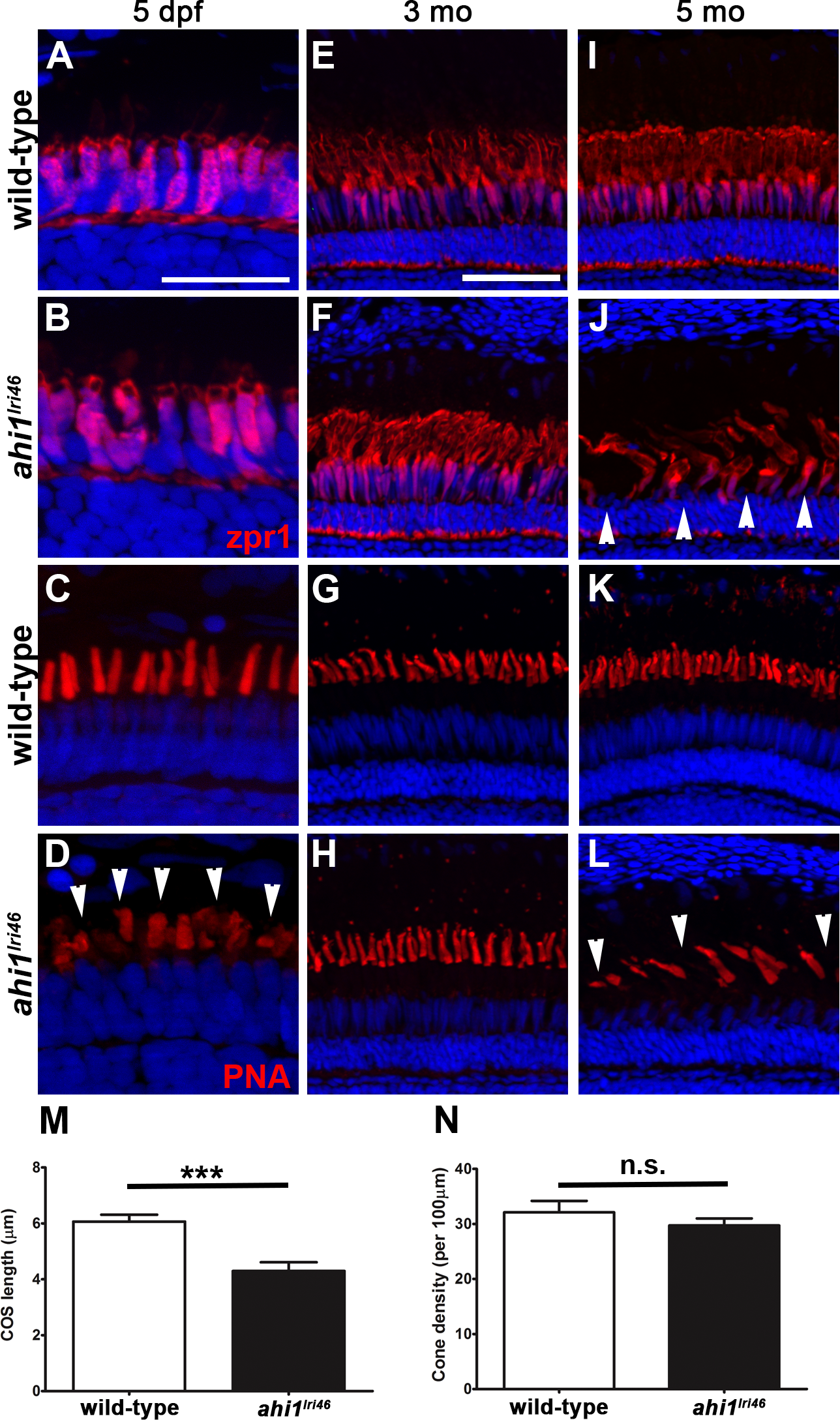
Fig. 7
Immunohistochemical analysis of cone photoreceptors in ahi1lri46 mutants. (A, B, E, F, I, J) Red-green double cones are labeled with zpr1 (red). Cone inner segments are preserved at 5 dpf and 3 months of age, but missing (arrowheads) at 5 months. (C, D, G, H, K, L) PNA staining (red) revealed that mutants had shorter cone outer segments ([D], arrowheads) at 5 dpf and missing at 5 months ([L], arrowheads). (M) Quantification of cone outer segment lengths between wild-type and ahi1lri46 mutants at 5 dpf. ***P < 0.001. N, quantification of cone density at 5 dpf.
Figure Data
Acknowledgments
This image is the copyrighted work of the attributed author or publisher, and
ZFIN has permission only to display this image to its users.
Additional permissions should be obtained from the applicable author or publisher of the image.
Full text @ Invest. Ophthalmol. Vis. Sci.