
Image
|
Figure Caption
Fig. 2
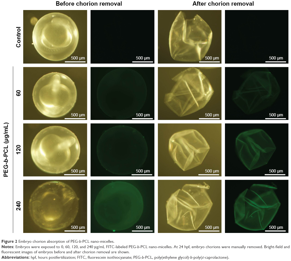
Figure 2 Embryo chorion absorption of PEG-b-PCL nano-micelles.
Notes: Embryos were exposed to 0, 60, 120, and 240 μg/mL FITC-labeled PEG-b-PCL nano-micelles. At 24 hpf, embryo chorions were manually removed. Bright-field and fluorescent images of embryos before and after chorion removal are shown.
Abbreviations: hpf, hours postfertilization; FITC, fluorescein isothiocyanate; PEG-b-PCL, poly(ethylene glycol)-b-poly(ε-caprolactone).
Acknowledgments
This image is the copyrighted work of the attributed author or publisher, and
ZFIN has permission only to display this image to its users.
Additional permissions should be obtained from the applicable author or publisher of the image.
Full text @ Int. J. Nanomedicine