
|
Fig. S3
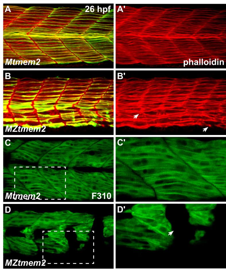
Disruption of fast muscle fiber attachment in MZtmem2 mutants. (A-D) Immunofluorescence reveals muscle fiber organization, using phalloidin (red in A,B) to recognize both fast and slow fibers, F59 (green in A,B) to recognize slow fibers (Devoto et al., 1996), and F310 (green in C,D) to recognize fast fibers (Nord et al., 2014); lateral views, dorsal up, at 26 hpf. (A,B) In addition to exhibiting detachment of F59+ slow muscle fibers (B); see also Fig. 1H), MZtmem2 mutants display detachment of F59- fast muscle fibers (arrows, B′), in contrast to the attached fibers observed in Mtmem2 siblings (A). (C,D) Similarly, MZtmem2 mutants (D) display detachment of F310+ fast muscle fibers (arrow, D2), in contrast to the attachment seen in Mtmem2 siblings (C). C′ and D′ show closer views of regions outlined by white rectangles in C and D.