
|
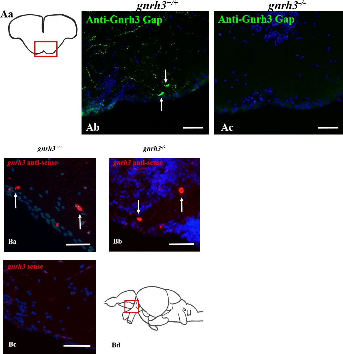
Fig. 3
gnrh3 mRNA but not Gnrh3 protein detectable in gnrh3-/- fish.
(A) Immunohistochemistry on adult coronal brain sections (Aa) using anti-zebrafish Gnrh3 Gap (green) demonstrates the presence of Gnrh3 signal in the form of somas (white arrows) and fibers in the gnrh3+/+ pre-optic area of the brain (Ab). However, no Gnrh3 signal was found in the gnrh3-/- pre-optic area (Ac) or in any other region of the brain. (B) In situ hybridization on adult sagittal brain sections (Bd) using gnrh3 DIG-labeled riboprobes. The anti-sense gnrh3 riboprobe demonstrated mRNA (red) in the ventral telencephalon and pre-optic area of both gnrh3+/+ (Ba) and gnrh3-/- (Bb) fish. The sense gnrh3 riboprobe demonstrated no gnrh3 mRNA signal in any brain regions in the gnrh3+/+ fish (Bc). Scale bars = 50 µm.