
|
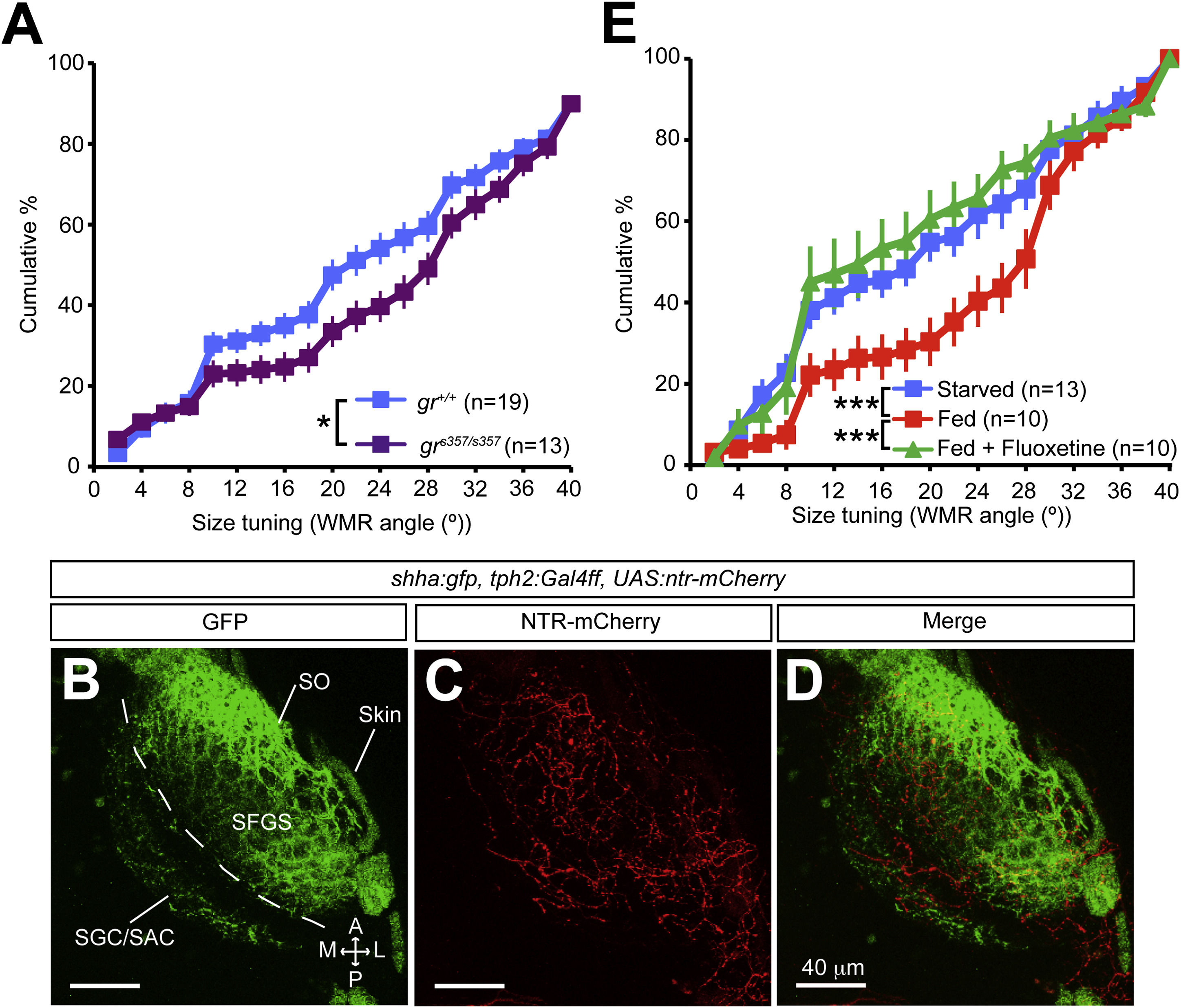
Fig. 7
The HPI Axis and the Serotonergic System Modulate Visual Information Processing in the Tectum
(A) Graph depicting cumulative percentages of WMR angles for PVNs in starved 7 dpf elavl3:Gal4, UAS:GCaMP5, grs357/s357 and starved control elavl3:Gal4, UAS:GCaMP5, gr+/+ larvae. p = 0.02, two-sample Kolmogorov-Smirnov test.
(B-D) Confocal images of a 7 dpf shha:gfp, tph2:Gal4ff, UAS:ntr-mCherry larva showing the presence of serotonergic innervation (red neurites in C and D) in the tectum neuropil (GFP-positive RGC axons in B and D). A, anterior; L, lateral; M: medial; NTR, nitroreductase; P, posterior; SAC, stratum album centrale; SFGS, stratum fibrosum et griseum superficiale; SGC, stratum griseum centrale; SO, stratum opticum.
(E) Graph comparing average cumulative percentages of WMR angles for PVNs of fed elavl3:Gal4, UAS:GCaMP5 larvae treated with 1.5 µM fluoxetine and untreated fed or starved controls. Fluoxetine abolished the satiety-induced change of PVN population response. ***p < 0.001, two-sample Kolmogorov-Smirnov tests. Data are presented as mean ± SEM.