
Image
|
Figure Caption
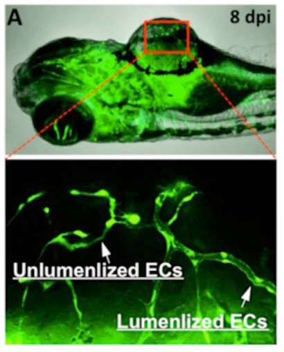
Fig. S2
Lumenization of solid angiogenic sprouts in xenografted microtumor on zebrafish.
(A) The coexistence of lumenized and unlumenized angiogenic circuits in xenografted tumors at 8 dpi (days post cell injection). (B) The formation of endothelial lumens via the dynamic fusion of intracellular vacuoles (arrows). (C) Schematic shows the process of endothelial cord lumenization in the xenografted tumor in zebrafish.
Acknowledgments
This image is the copyrighted work of the attributed author or publisher, and
ZFIN has permission only to display this image to its users.
Additional permissions should be obtained from the applicable author or publisher of the image.
Full text @ Sci. Rep.