
|
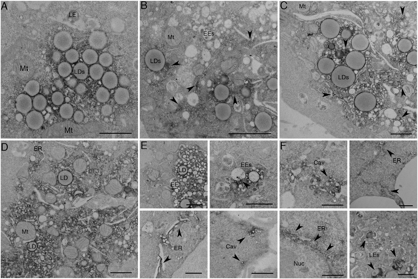
Fig. 2
APEX-GBP Allows for Time-Resolved Protein Redistribution
(A–F) BHK cells were transfected and serum starved for (A) 0 min, (B) 30 min, (C) 1 hr, (D) 2 hr, (E) 4 hr, and (F) 8 hr to track the redistribution of GFP-CavDGV by APEX-GBP in a time-resolved manner. Arrows highlight regions where electron density has re-distributed from lipid droplets to other intracellular organelles. LD, lipid droplet; Mt, mitochondria; EE, early endosome; LE, late endosome; ER, endoplasmic reticulum; Nuc, Nucleus; Cav, Caveolae. Scale bars represent 500 nm. The above localizations were confirmed by confocal microscopy; see Figure S2.
Reprinted from Developmental Cell, 35(4), Ariotti, N., Hall, T.E., Rae, J., Ferguson, C., McMahon, K.A., Martel, N., Webb, R.E., Webb, R.I., Teasdale, R.D., Parton, R.G., Modular Detection of GFP-Labeled Proteins for Rapid Screening by Electron Microscopy in Cells and Organisms, 513-25, Copyright (2015) with permission from Elsevier. Full text @ Dev. Cell