
Fig. 7
- ID
- ZDB-IMAGE-151228-18
- Genes
- Antibodies
- Source
- Figures for Mendieta-Serrano et al., 2015
|
Fig. 7
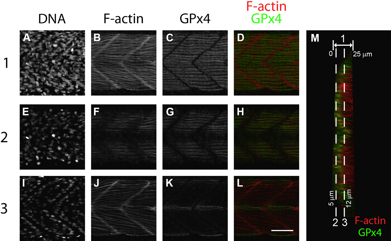
GPx4b is localized in the slow muscle fibers 24 hpf zebrafish embryos myotomes. To identify in detail the GPx4b positive tissue in myotomes, different maximum intensity projections were generated from the confocal z optical slices. 1 (A to D), maximum intensity projections of full z stacks, representing approximately 25 µm. 2 (E to H), maximum intensity partial projections of slices located 5 µm depth shows that slow muscle fibers are located closer to the embryo surface, and the pattern of F-actin staining shows that slow muscle fibers are thicker and parallel to the main embryo axis (F). The GPx4b staining in slow muscle fibers is intense (G). 3 (I to L), deeper muscle fibers, at approximately 12 µm, show a different F-actin staining pattern, presenting thinner fibers (J) and a decreased GPx4 signal (K). Hoechst-stained embryos (A, E, and I). F-actin, phalloidin Alexa 488-stained embryos (B, F and J). GPx4b immunolocalization (C, G and K). Overlays (D, H and L), superimposition of F-actin (in red) and GPx4 (in green). M, orthogonal slice. Scale bar 50 µm.
Reprinted from Gene expression patterns : GEP, 19(1-2), Mendieta-Serrano, M.A., Schnabel-Peraza, D., Lomelí, H., Salas-Vidal, E., Spatial and temporal expression of zebrafish glutathione peroxidase 4 a and b genes during early embryo development, 98-107, Copyright (2015) with permission from Elsevier. Full text @ Gene Expr. Patterns