
Image
|
Figure Caption
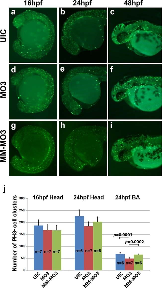
Fig. 4
Cell proliferation is unchanged in MO3 injected embryos. Whole mount immunohistochemistry with mitotic marker anti-phosphohistone H3 shows proliferating cells in developing embryos. UIC (a–c), MO3 (d–f), and control MM-MO3-injected embryos (g–i) embryos at 16 hpf (a, d, g), 24 hpf (b, e, h), and 48 hpf (c, f, i). (j) Quantification of number of proliferating cells in UIC, MO3, and control MM-MO3 injected embryos at 16 and 24 hpf.
Figure Data
Acknowledgments
This image is the copyrighted work of the attributed author or publisher, and
ZFIN has permission only to display this image to its users.
Additional permissions should be obtained from the applicable author or publisher of the image.
Full text @ Genesis