
Image
|
Figure Caption
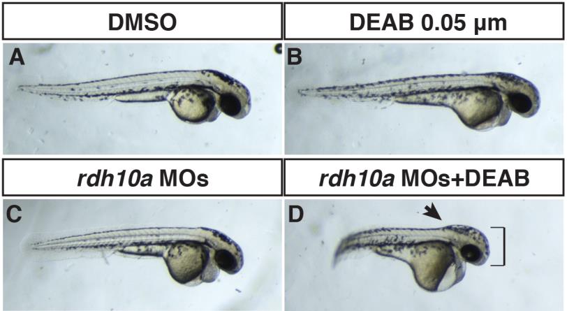
Fig. S3 Rdh10a depletion enhances defects caused by inhibition of Aldh1a2.
(A) Control sibling embryo treated with DMSO. (B) Embryo treated with 0.05 µM DEAB (a suboptimal concentration) that has mild defects indicated of loss of RA signaling. (C) Rdh10a deficient embryo. (D) Rdh10a deficient embryos treated with the suboptimal concentration of DEAB results in an interaction that produces defects reminiscent of Rdh10a depleted nls mutant embryos (Fig 4). The head is enlarged (brackets) and the border between the hindbrain and anterior spinal cord is accentuated (arrow), indicating the hindbrain is anteriorized.
Acknowledgments
This image is the copyrighted work of the attributed author or publisher, and
ZFIN has permission only to display this image to its users.
Additional permissions should be obtained from the applicable author or publisher of the image.
Full text @ PLoS One