
|
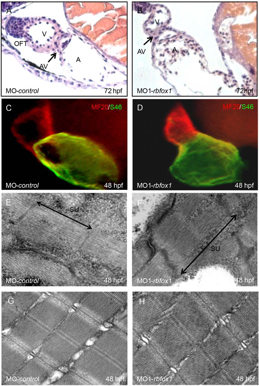
Fig. 3
rbfox1 deficency does not influence heart morphology. (A,B) H&E-stained sagittal histological sections of MO-control and MO1-rbfox1 morphant hearts at 72hpf. Morphants display normal heart morphology with distinct endocardial and myocardial cell layers in the atrium (A) and ventricle (V), and a clear differentiation and demarcation of the atrium and ventricle by the atrio-ventricular ring (AV). OFT, outflow tract. (C,D) Atrial- and ventricle-specific myosin heavy chains are expressed normally, also suggesting that there is normal molecular chamber specification [green, antibody against atrial-specific myosin (S46); red, antibody against ventricular and atrial myosin (MF20)]. (E–H) Ultrastructural analysis of heart and skeletal muscle cells of MO-control- and MO1-rbfox1-injected embryos at 48hpf showing organized sarcomere units (SU) with thin and thick myofilaments in well-aligned bundles and discernible AI-, M- and Z-bands.