
|
Fig. 1
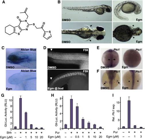
Eggmanone Affects Embryonic Zebrafish Patterning through Inhibition of Hedgehog Signaling
(A) The chemical structure of eggmanone (Egm), (3-(2-methylallyl)-2-((2-oxo-2-(thiophen-2-yl)ethyl)thio)-5,6,7,8-tetrahydrobenzo[4,5]thieno[2,3-d]pyrimidine-4(3H)-one), is shown.
(B) Zebrafish embryos treated with 2 µM Egm starting at 4 hr post fertilization (hrpf) exhibited a range of phenotypes found in Hh pathway mutants, including ventral tail curvature, loss of pectoral fins, and smaller eyes.
(C) Alcian blue staining following 2 µM Egm treatment at 4 hrpf revealed altered craniofacial development, including the jaw.
(D) Trunk slow muscles immunostained with anti-MyHC antibody (F59) showed altered slow-muscle formation upon Egm treatment (2 µM).
(E) Egm treatment (1 µM) abolished Hh-responsive Ptc1 expression in adaxial cells at 12 hrpf (arrows).
(F) Egm treatment (1 µM) ablated Hh-responsive Ptc1 expression in the pectoral fin bud at 48 hrpf (arrows and asterisks).
(G) Egm inhibited Sonic hedgehog (Shh)-responsive Gli-luciferase (Gli-Luc) reporter activity in a dose-dependent manner when stimulated with Shh-conditioned medium (n = 4 for each condition, results represented as mean relative luciferase units (RLU) ± SEM; p value < 0.0184, starting at 1 µM).
(H) Egm inhibited purmorphamine-induced (3 µM) Gli-Luc reporter activity in a dose-dependent manner (mean ± SEM, n = 4 for each condition; p value < 0.0054, starting at 0.5 µM).
(I) Egm inhibited purmorphamine-induced (3 µM) Ptc1 expression in NIH 3T3 fibroblasts (mean ± SEM, n = 3, expression normalized to GAPDH; p value < 0.003, starting at 1 µM). Related to Figures S1 and S3.