
|
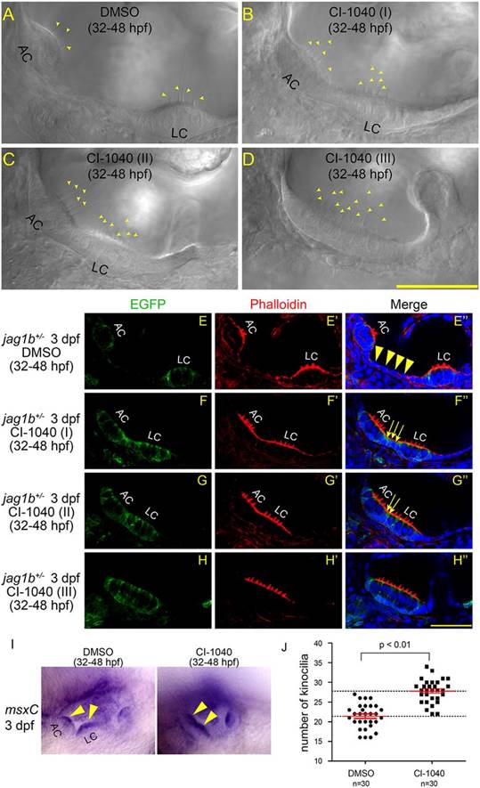
Fig. 7
Inhibition of MAPK signaling generates a larger crista. (A-D) DIC confocal imaging of CI-1040-treated jag1b+/- embryos at 3dpf. Kinocilia are indicated by arrowheads. AC, anterior crista; LC, lateral crista. (E-H′′) Phalloidin staining of MAPKK inhibitor CI-1040-treated jag1b+/- embryos at 3dpf. Lateral views. Anterior towards the left and dorsal upwards. Scale bars: 40µm. Arrows and arrowheads indicate cells between AC and LC (E′′-G′′). (I) Whole-mount in situ hybridization of DMSO-and CI-1040-treated embryos. The probes used is msxC antisense. Arrowheads indicate cristae. (J) Total kinocilia number in the anterior and lateral cristae of DMSO-treated embryos and kinocilia number in the fused crista of CI-1040-treated embryos. Data are shown as mean±s.e.m. Statistical analysis was performed using a t-test. n indicates the number of inner ears for each experimental group.