
|
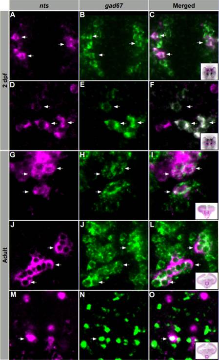
Fig. 3 NTS neurons are primarily GABAergic. Confocal imaging of double fluorescent ISH experiments in larvae and adult zebrafish. A–F: 2 dpf larvae showing colocalization of nts- (magenta) and gad67- (green) expressing cell bodies within the PO (A–C) and hypothalamus (D–F). G–O: Transversal adult brain sections showing colocalization of nts- (magenta) and gad67- (green) expressing cell bodies within the PM (G–I), TPp (J–L), and Hc (M–O). Arrows indicate representative coexpressing cells (white). All images are single confocal sections. PO, preoptic region; H, hypothalamus; PM, magnocellular preoptic nucleus; TPp, periventricular nucleus of the posterior tuberculum; Hc, caudal zone of the periventricular hypothalamus.