
|
Fig. 5
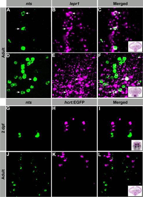
LepR1-expressing NTS neurons are located adjacent to HCRT neurons in the hypothalamus. Confocal imaging of double fluorescent ISH (A–F) and ISH-immunohistochemistry (G–L) experiments in larvae and adult zebrafish. A–F. Transversal adult brain sections showing colocalization of nts- (green) and lepR1- (magenta) expressing cell bodies within the Hv (A–C) and Hc (D–F). G–L: 2 dpf larvae (G–I) and transversal adult brain sections (J–L) showing nts- (green) and hcrt- (magenta) expressing neurons (within the PO and Hv, respectively) that do not colocalize. Arrows indicate representative coexpressing cells (white). All images are single confocal sections. PO, preoptic region; Hv, ventral zone of the periventricular hypothalamus; Hc, caudal zone of the periventricular hypothalamus.