
|
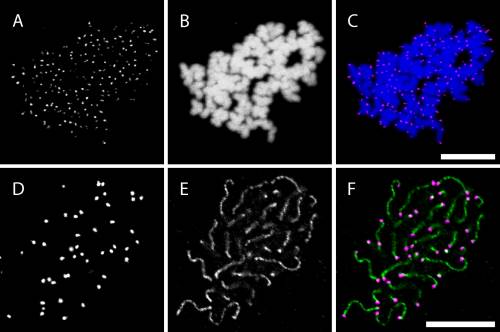
Fig. 1
FISH using a Cy3-conjugated (CCCTAA)3 PNA telomere probe in metaphase cells and pachytene spermatocytes. A–C: FISH using a telomere probe in metaphase chromosomes prepared from the cells of a 1-day-old zebrafish embryo. Panel A shows FISH signals, panel B shows nuclear staining with TO-PRO-3, and panel C shows a merged image of panels A and B. More than 90% of telomeres were clearly detected. D–F: FISH using a telomere probe in combination with immunocytochemical labeling of Sycp3 in pachytene spermatocyte spreads. Panel D shows FISH signals, panel E shows Sycp3 signals, and panel F shows a merged image of panels D and E. Telomere signals were detected at both ends of Sycp3-positive structures. Scale bars = 10 µm.