
|
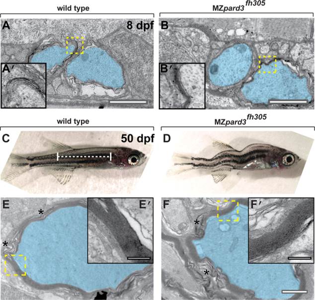
Fig. 7 pard3 is not essential for Schwann cell myelination. A,B: Transmission electron micrographs of coronal sections through the trunk region of 8 dpf wild-type (A) and MZpard3fh305 mutant (B) larvae. Motor axons are pseudocolored blue. Areas indicated by dashed boxes are shown at higher magnification in insets. Multiple layers of myelin membrane are evident in both wild-type and mutant. C,D: Lateral views of 50 dpf wild-type (C) and MZpard3fh305 mutant (D) zebrafish. Dashed white line indicates region of coronal sections obtained for electron microscopy. MZpard3fh305 mutants are shortened and display severe body deformation, manifesting as a variable curved body axis. E,F: Transmission electron micrographs of coronal sections through the trunk region of 50 dpf fish. Areas indicated by dashed boxes are shown at higher magnification in insets. Myelin ultrastructure is similar in wild-type (E,E′) and MZpard3fh305 mutant (F,F′). Scale bars = 1 µM in A-D; 0.25 µM in A′,B′,C′,D′.