
|
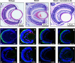
Fig. 8
Effect of RIP signalling inhibitors on pde6cw59 mutant phenotype at 7 d.p.f. Retinal histology in (a) wild-type (wt), (b) untreated mutant (mt) and (c) mt retina from embryos treated with 100mM necrostatin-1s (N). White arrows in a–c indicate the photoreceptor nuclear layer of the retina. Rip1 labelling (red) in wt retina (d), in mt retina co-localizing with EGFP-expressing cones that are dying (e), and in mt retina treated with necrostatin-1s (f). (g) rip1 labelling in the retina of pde6cw59 mutant embryos treated with 20μM necrosulfonamide (S). TUNEL labelling in non-transgenic pde6cw59 mutant embryos (h–k) corresponding to the treatment panels (d–g) above. Scale bar in d=20μm