
|
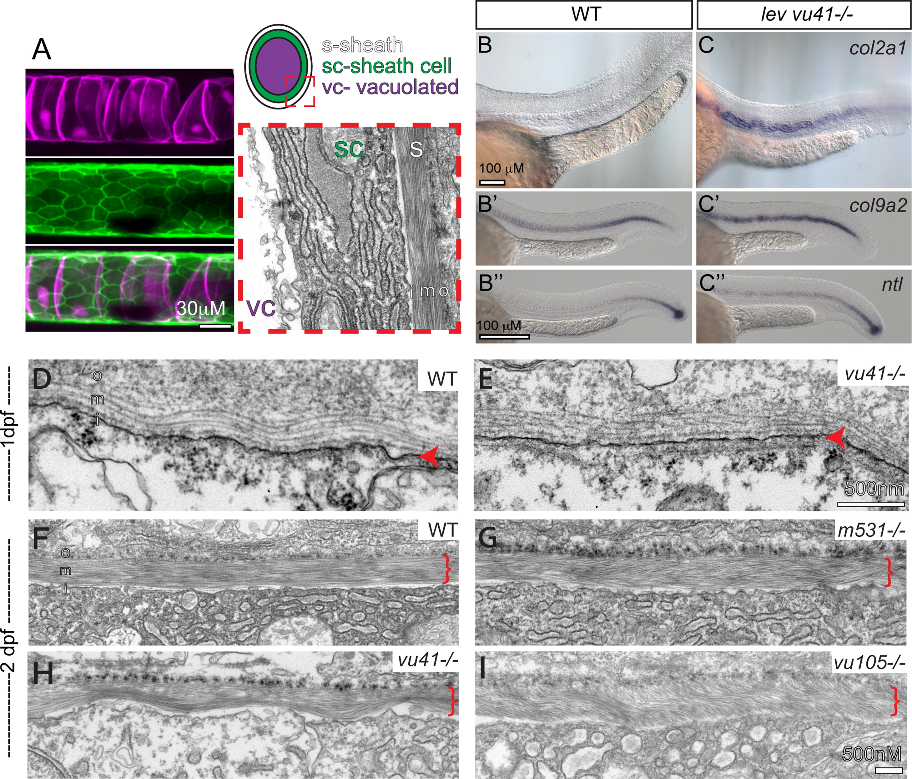
Fig. 2 col8a1a mutations disrupt the normal morphology of the notochord sheath. (A) The primary components of the notochord tissue at embryonic maturation illustrated with a confocal (A, B, and C), cartoon (D), and transmission electron micrographs (TEM) (E). The large vacuolated cell layer (VC; Tg(TK5xC twhh:memRFP) expression; magenta), surrounded by a sheath epidermis (SC; 1.75KB col2a1 GFP-CAAX; green) are shown in a lateral view with a confocal projection. The cartoon and the TEM image are transverse views highlighting the extracellular tri-laminar notochord sheath (S) composed of laminin-, collagen- and elastin-rich components. (B–C′′) Representative examples of in situ hybridization of chordamesodermal genes col2a1 (B and C), col9a2 (B′ and C′), and ntl (B3, C3) in WT (B–B′′) and levvu41 mutant (C–C′) embryos. (C) The expression of col9a2 and ntl transcripts is upregulated, especially in more anterior regions of the notochord (C′ and C′′) as compared to time matched sibling embryos (B′ and B′). TEM of transverse sections from WT and mutant embryos at 2dpf (D–I). (D) WT notochord at 1 dpf. (E) levvu41-/- notochord at 1dpf. (F) WT notochord sheath at 2dpf display very straight, well organized medial sheath layer. Extracellular notochord sheaths of lev m531-/- (G), levvu41-/- (H), and lev vu105-/- (I) mutants exhibiting a wavy, disordered organization of the medial layer at 2dpf. I – inner laminin-rich layer, m – medial, and o – outer collagen-rich layers of the extracellular sheath; SC – sheath cell, S – extra-cellular sheath, and VC – vacuolated cell.
Reprinted from Developmental Biology, 386(1), Gray, R.S., Wilm, T.P., Smith, J., Bagnat, M., Dale, R.M., Topczewski, J., Johnson, S.L., and Solnica-Krezel, L., Loss of col8a1a function during zebrafish embryogenesis results in congenital vertebral malformations, 72-85, Copyright (2014) with permission from Elsevier. Full text @ Dev. Biol.