
|
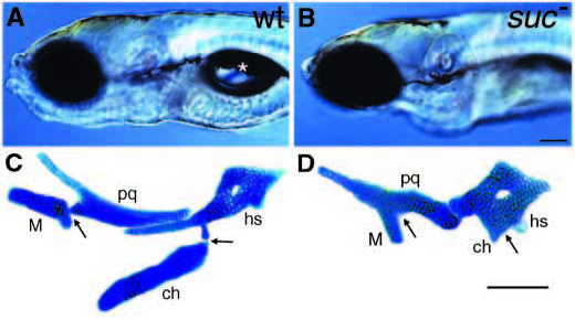
Fig. 1 suc mutants have reduced ventral cartilages. (A,B) Lateral views of live wild-type (A) and sucker mutant (B) larvae at 4 days. Mutants lack lower jaws and do not form swim bladders (asterisk in A). (C,D) Alcian-stained cartilages from a wild type (C) and suc mutant (D) dissected from the arches and flat-mounted. In wild types, dorsal and ventral elements in the first arch (pq, palatoquadrate; M, Meckel’s) and second arch (hs, hyosymplectic; ch, ceratohyal) are separated by joints (arrows). In mutants, ventral cartilages are reduced and fused to dorsal cartilages at what we interpret to be the sites of the missing joints (arrows). Scale bars: 100 μm.