
|
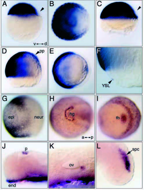
Fig. 4 bmp7 expression during embryogenesis. (A) 30% epiboly stage (lateral view). Expression extends throughout the blastoderm, except its dorsal marginal aspect (arrowhead). Animal pole (B) and lateral (C) views of a germ ring stage embryo showing bmp7 downregulation in the dorsal quadrant. (D-F) 60% epiboly stage. Lateral (D) and transverse optical cross-sectional (E) views showing expression throughout the ventral gastrula and in the prechordal plate (pp) (D). (F) Close-up of the ventral margin showing expression in the YSL. (G) Lateral view at 90% epiboly. Robust expression is seen at the border between epidermal (epi) and neurogenic (neur) ectoderm and in the ventral margin. Faint expression is observed throughout the epidermal territory. (H,I) 3-somite stage. (H) Dorsal cephalic view. Transcripts accumulate at the border of the hatching gland (hg). Faint expression is detected outside the neural plate. (I) Posterior view. Transcripts are localized to the tail bud (tb) and the border between neural and non-neural posterior ectoderm. At 24 h.p.f. expression is observed in (J) the endoderm (end) and the pineal gland (p), (K) the posterior aspect of the otic vesicle (ov) and (L) dorsal posterior neural tissue (spc) of the tail. A-G, dorsal to the right; H-K, anterior to the left.