
Image
|
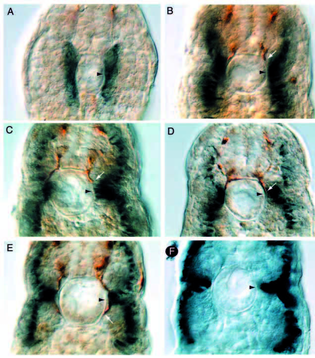
Figure Caption
Fig. 5 Motor growth cone and adaxial cell migration coincide. (A-E) Cross-section of somitic segments (18-12) of wild-type embryos at 20 hpf stained with znp-1 antibody (brown) and F59 antibody (blue). White arrows point to the progressing growth cone and arrowheads point to distal end of the common path in the region of the future horizontal myoseptum. (F) Cross-section of somitic segment 14 of a diwanka mutant embryo at 24 hpf stained with F59 (blue), demonstrating that, in diwanka mutant embryos, the migrated adaxial cells are indistinguishable from those in wild-type siblings.
Acknowledgments
This image is the copyrighted work of the attributed author or publisher, and
ZFIN has permission only to display this image to its users.
Additional permissions should be obtained from the applicable author or publisher of the image.
Full text @ Development