
|
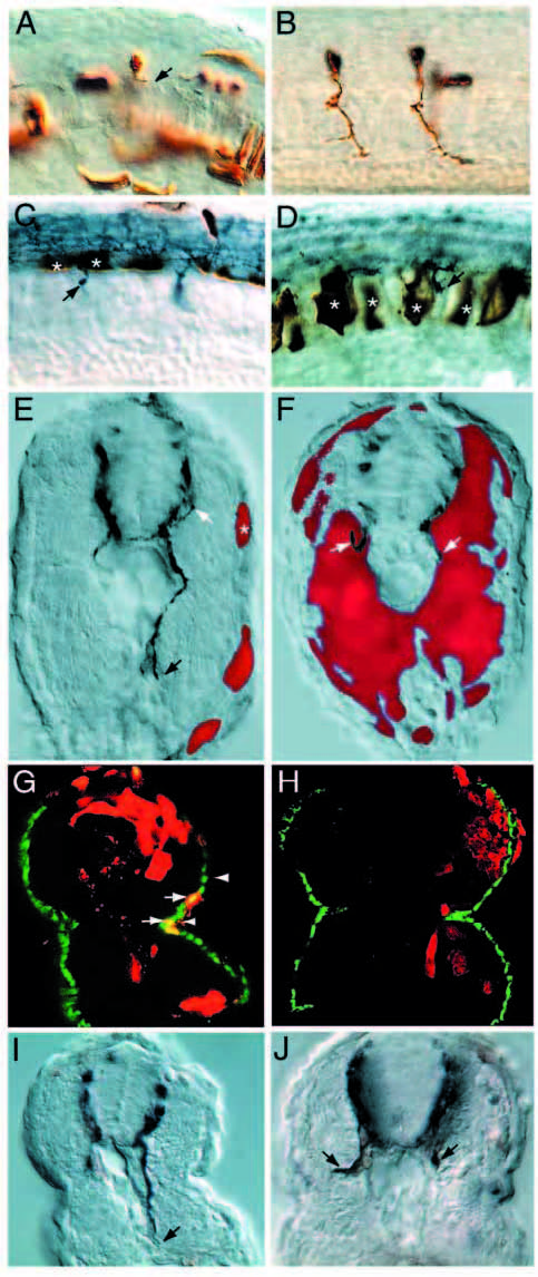
Fig. 4 Analysis of chimeric mosaic embryos reveals that diwanka activity is required in adaxial cells. (A) A genotypically wild-type motoneuron (brown) which developed in diwanka mutant embryos and failed to exit the spinal cord (arrow). (B) Two genotypically mutant CaP motoneurons (brown) which developed in wild-type embryos and displayed normal axonal trajectories. (C,D) Wild-typederived floorplate (asterisks in C) or notochord cells (asterisks in D) failed to restore motor axonal trajectories. Arrows point to znp-1- positive motor growth cones, which failed to complete the common path. (E,F) Cross-section of mosaic diwanka embryos showing wildtype- derived cells (in red) and znp-1-positive motor axons in dark blue. (E) Small clones of genotypically wild-type cells in the lateral portion of the somite (asterisk) restored the motor axon projections of CaP (black arrow) and MiP (white arrow, only partially in focal plane). (F) In contrast, large clones of genotypically wild-type cells in the medial part of the somites failed to restore motor axon projections. (G) Cross-section showing colocalization between wildtype- derived cells (red) able to rescue the axonal phenotype in the right hemisegment and F59-positive adaxial cells (green). Arrows point to double-labeled cells (yellow), located between the lower end of the spinal cord (upper arrowhead) and the horizontal myoseptum (lower arrowhead). The rescued motor axon is shown in Fig. 4I. (H) Cross-section through a segment in which wild-type-derived cells (red) occupied most of the segment, but F59-positive adaxial cells (green) are of diwanka mutant origin. There is no colocalization between wild-type-derived cells (red) and F59-positive adaxial cells (green) and the motor trajectories were not rescued (see Fig. 4J). (I) DIC image of the cross-section shown in G. The arrow points to the rescued, znp-1-positive motor axon. (J) DIC image of the crosssection shown in H. Arrows point to mutant, znp-1-positive motor axons.