
|
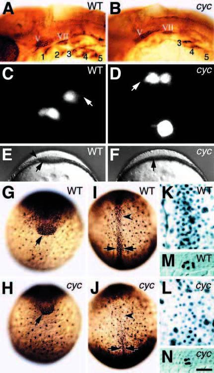
Fig. 8 Embryos mutant for cyclops have fewer dorsal endodermal and mesodermal precursors. (A,B) 30 hour embryos, side view, labeled with Zn8 antibody. The antibody labels the cell surfaces of the pharyngeal pouches (indicated by numerals 1-5) and sensory neurons in the cranial ganglia. (C,D) 100% epiboly, dorsal view; endodermal precursor cells in the prechordal plate of live embryos appear similar in both wild-type and mutant; arrows, filopodia. The very round and intensely bright cell in the lower field in D is preparing to divide and did so immediately after this image was captured. Progeny of both clones later gave rise to pharyngeal endoderm. (E,F) 90% epiboly, front view; the prechordal plate (arrow) is noticeably thinner in mutant embryos, and lacks a visible lateral boundary (arrowhead). (G-J) 100% epiboly stage embryos labeled with anti-Fkd2 illustrating the anterior prechordal plate (arrow); the posterior prechordal plate (arrowhead); and the notochord (between arrows). (K,L) Higher magnification of embryos (I,J) showing the endodermal precursors in a deep plane of focus. The boundary between prechordal plate endoderm and notochord, obvious in the cyclops embryo, is towards the lower field. (M,N) 100% epiboly stage embryos labeled with anti-Ntl, a notochord-specific marker. Shown is a transverse section (10 μm) mid-trunk through the embryo. Using a 40x lens, we counted all the intensely labeled Fkd2-positive nuclei in the anterior prechordal plate (wt: median = 195, n=3; cyclops: median = 106, n=3). Similarly, we counted all labeled nuclei in a 100x100 μm field of the posterior prechordal plate (wt: median = 409, n=3; cyclops: median = 210, n=3) and the notochord (wt: median = 217, n=3; cyclops: median = 120, n=3). Endodermal precursors, labeled nuclei adjacent to the yolk, comprise a subset of the prechordal plate field (wt: median = 127, n=3; cyclops: median = 60, n=3). Scale bar: 100 μm (A,B, E,F and G-J), 25 μm (K-N), 8 μm (C,D); abbreviations: V, trigeminal ganglion; VII, facial ganglion.