
|
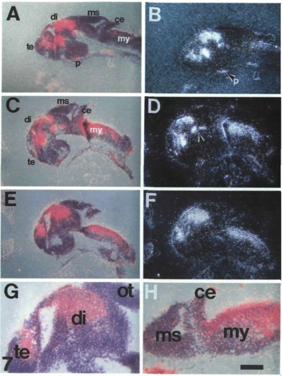
Fig. 7 pax-6 is expressed in distinct regions at 32 and 36 h. Parasagittal sections of 32 h (A, B) and 36 h (C - H) fish were hybridized with a pax-6 probe (probe A, probe C gave identical results). Double-exposures of brightfield (blue) and dark-field (red) images (A, C, E) and dark-field images (B, D, F) of the same sections. Expression is detectable in the myelencephalon up to the border between the myelencephalon and the cerebellum (A - F, H). The density of grains over the cerebellum and the mesencephalon is the same as that seen throughout the slide and do not represent specific hybridization to pax-6 transcripts, pax-6 is expressed in the diencephalon and the telencephalon (A - F, G) and specific subareas within these regions (C, D: arrow, G). Abbreviations: ce, cerebellum; di, diencephalon; ms, mesencephalon; my: myelencephalon; ot: optic tectum; p, pituitary gland; te, telencephalon. Anterior is to the left, dorsal up. Scale bar is 100 μn in A - F and 40 μm in G, H.