
|
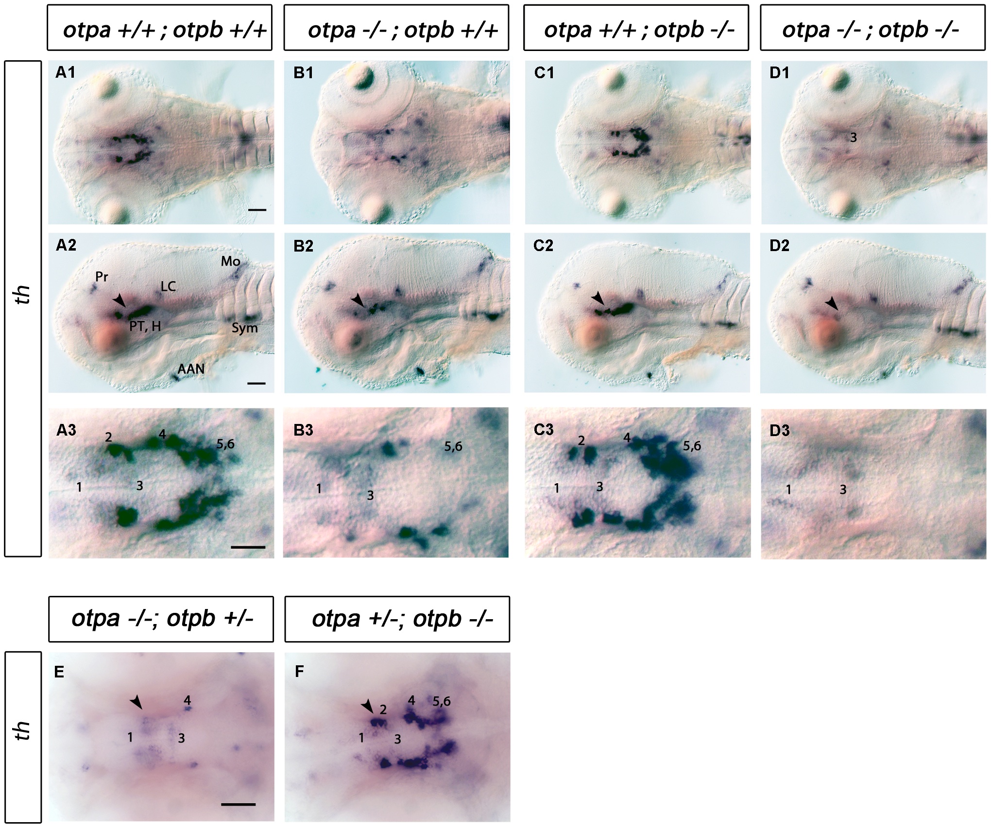
Fig. 3 Analysis of DA neurons by expression of th in otpa and otpb single and double mutant larvae.
(A–D) Whole-mount in situ hybridization of 3 dpf larvae reveals reduction of th expression in the posterior tuberculum of otpa and total loss of the expression in otpa;otpb double mutants (arrowhead). Other th expressing domains are not affected. (A1–D1, A3–D3) Dorsal views, anterior at left; (A2–D2) lateral views, dorsal up. Scale bar is 50 μm. (E,F) Whole-mount in situ hybridization of 3 dpf larvae reveals reduction of th expression in the posterior tuberculum of otpa mutant, otpb heterozygous larvae (E) (arrowhead). No clear reduction is detected in the posterior tuberculum of otpb mutant, otpa heterozygous larvae (F) (arrowhead). Dorsal view, anterior at left. Scale bar is 50 μm. Abbreviations: AAC, arch associated cluster; DC, diencephalic cluster; H, hypothalamus; LC, locus coeruleus; MO, medulla oblongata; Pr, pretectum; PT, posterior tuberculum. Numbers indicate dopaminergic neurons in the ventral thalamic cluster (1) and posterior tuberculum/hypothalamus (2–6) according to [20]. Scale bar is 50 μm.