
Image
|
Figure Caption
Fig. 4
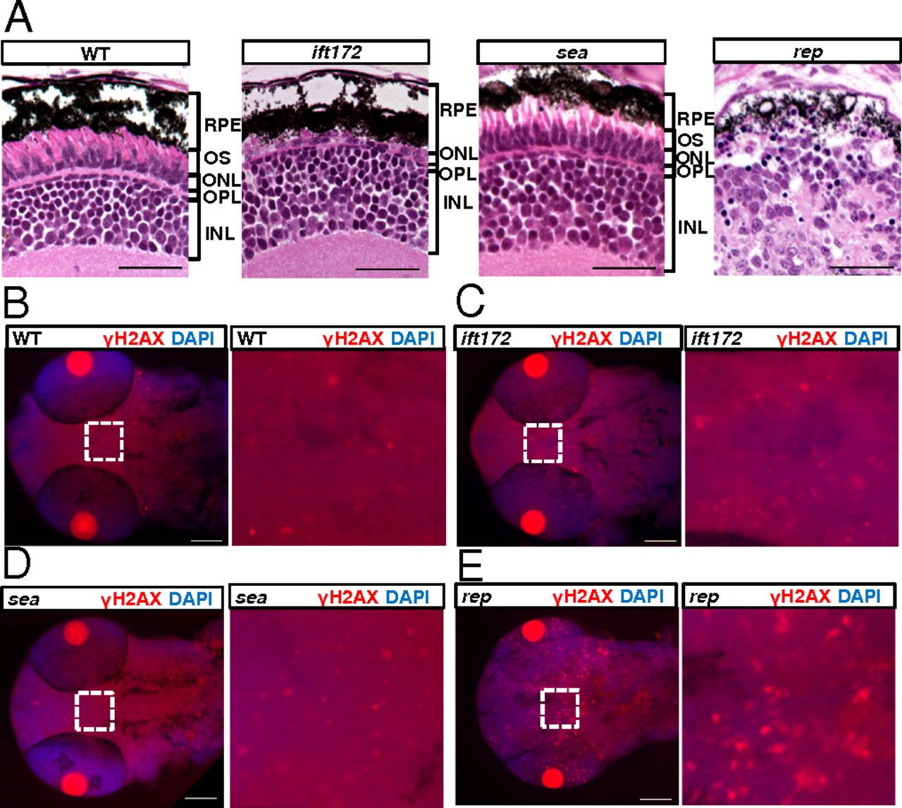
reptinhi2394 mutants show cilia-independent phenotypes. (A) Sections of the eye at 5 dpf. RPE, retinal pigment epithelium; OS, outer segment; ONL, outer nuclear layer; OPL, outer plexiform layer; INL, inner nuclear layer. (Scale bar, 20 μm.) (B–E) The head and eye region at 2-dpf mutants stained with an anti-γH2AX antibody in red. The boxed regions (Left) of B–E are magnified on corresponding Right. The pair of bright red circles in each image is the pair of lenses. (Scale bar, 100 μm.) WT, wild type; ift172, ift172hi2211 mutant; sea, seahorsehi3308 mutant; rep, reptinhi2394 mutant.
Figure Data
Acknowledgments
This image is the copyrighted work of the attributed author or publisher, and
ZFIN has permission only to display this image to its users.
Additional permissions should be obtained from the applicable author or publisher of the image.
Full text @ Proc. Natl. Acad. Sci. USA