
Image
|
Figure Caption
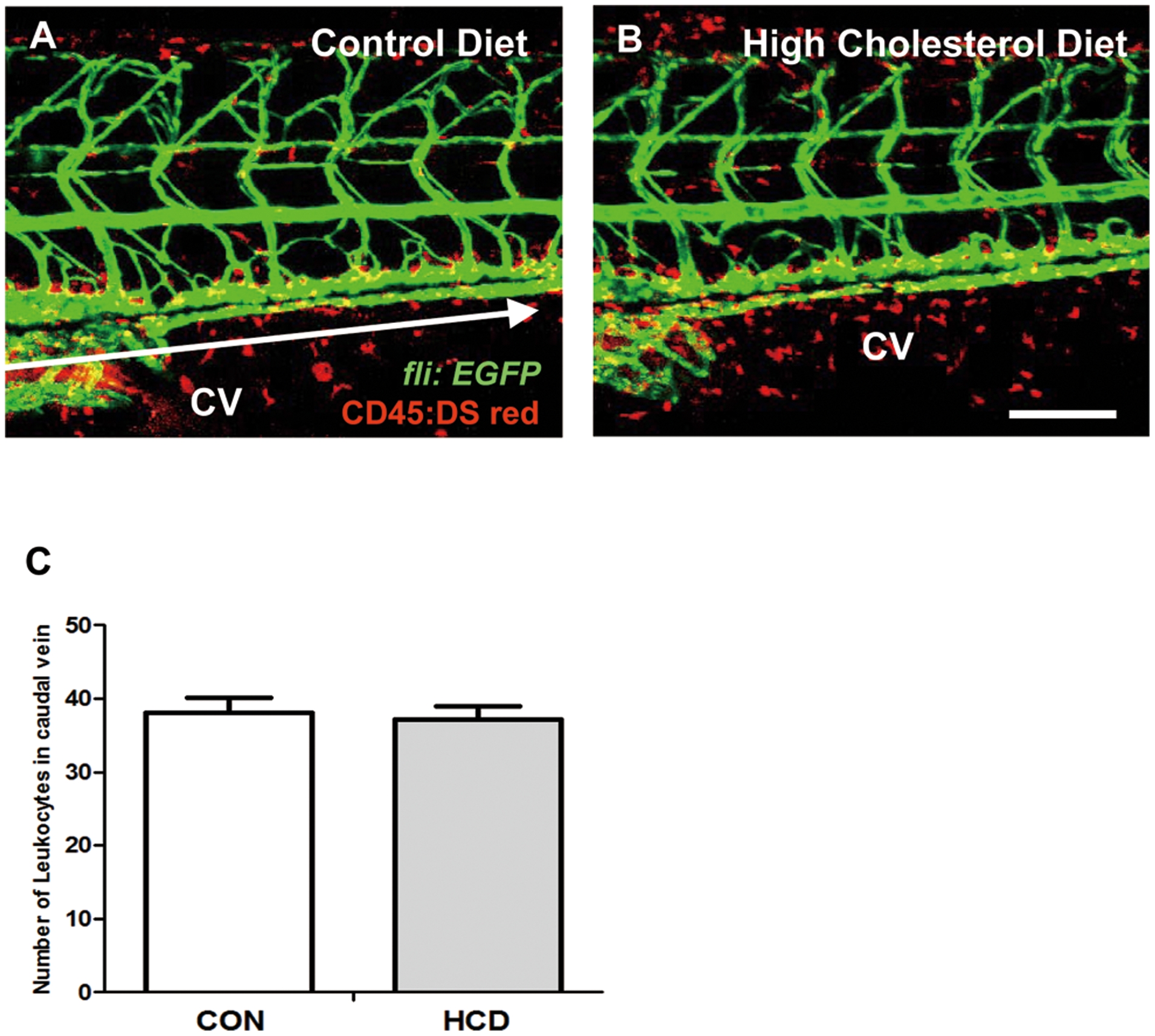
Fig. 2
Vascular leukocytes accumulation in HCD fed zebrafish. zebrafish larvae were fed a normal (control) diet (A) or a 4% cholesterol-enriched (HCD) (B) for 10 days.
Images of the caudal vasculature in live larvae show leukocytes accumulation in blood vessel or around tissues deposits in zebrafish larvae. Scale bar = 80 μm. The number of leukocytes in caudal vein (yellow) between each edge of arrow were counted in each a control diet and HCD group (each group n = 9) (C). CV, caudal vein.
Acknowledgments
This image is the copyrighted work of the attributed author or publisher, and
ZFIN has permission only to display this image to its users.
Additional permissions should be obtained from the applicable author or publisher of the image.
Full text @ PLoS One