
|
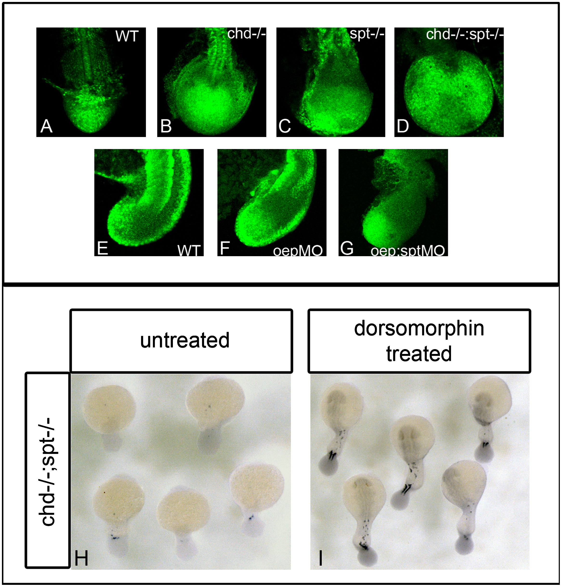
Fig. 3 BMP inhibition is required for cells to exit the tailbud and form somites. P-smad 1/5/8 antibody was used to detect BMP activity in the tailbud of embryos during somitiogenesis. (A)–(D) dorsal view of P-smad 1/5/8 antibody fluorescence in 17 hpf (16 som) embryos; (E)–(G) are lateral views of P-smad 1/5/8 staining in 17 hpf embryos. WT embryos have normal levels of active BMP ((A) and (E)). Chd-/- (B) and oepMO (F) embryos have elevated levels of BMP, possibly due to the roles of chd and oep in BMP inhibition. chd-/-;spt-/- (D) and oepMO;sptMO (G) embryos also have higher levels of BMP in the tailbud, however, elevated levels are found in only some areas of the tailbud. Elevated Bmp activity and lack of spt leads to phenotypes where progenitor cells cannot exit the tailbud to form somites. (H)–(I), dorsal view of myoD expression in 24 hpf embryos: chd;spt embryos treated with dorsomorphin, a small molecule inhibitor of BMP, at 12 hpf were partially rescued and able to produce tail somites. (23/25 had 5–9 somites) (I), whereas untreated chd;spt embryos do not produce somites (1/26 had 5–9 somites) (H).
Reprinted from Developmental Biology, 375(2), O'Neill, K., and Thorpe, C., BMP signaling and spadetail regulate exit of muscle precursors from the zebrafish tailbud, 117-127, Copyright (2013) with permission from Elsevier. Full text @ Dev. Biol.