
|
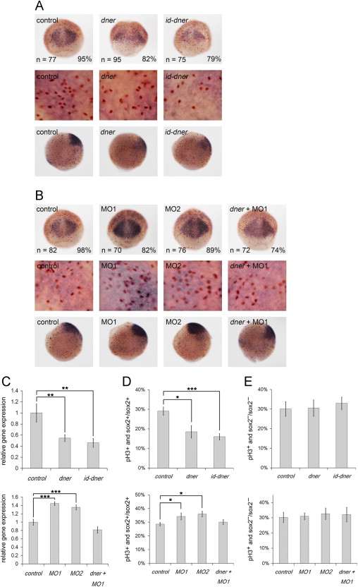
Fig. 2 Dner inhibits the proliferation of neural progenitors. Proliferation of neural progenitors was detected by in situ hybridization using a sox2 riboprobe (purple) and counterstaining with phosphohistone H3 antibody (brown) at 75% epiboly. (A–B) Top and middle panels are dorsal and bottom panels are lateral views. Middle panels show enlarged images of the corresponding top panels. (A) The proliferation of neural progenitors was reduced in dner- or id-dner-injected embryos compared with the controls. (B) Injection with either MO1 or MO2 induced the proliferation of neural progenitors, and the phenotypes caused by morpholino injection could be rescued by concomitant injection with dner cRNA. (C) The results of in situ hybridization were confirmed quantitatively by qPCR analysis. (D) The proportions of phosphohistone H3- and sox2-positive cells among the total sox2-positive cells were quantified. (E) The proportions of phosphohistone H3-positive and sox2-negative cells in sox2-negative cells counted in adjacent surface ectoderm showed no significant deviation in Dner-distorted embryos. N, p<0.05; NN, p<0.01; NNN, p<0.001.
Reprinted from Developmental Biology, 375(1), Hsieh, F.Y., Ma, T.L., Shih, H.Y., Lin, S.J., Huang, C.W., Wang, H.Y., and Cheng, Y.C., Dner inhibits neural progenitor proliferation and induces neuronal and glial differentiation in zebrafish, 1-12, Copyright (2013) with permission from Elsevier. Full text @ Dev. Biol.