
|
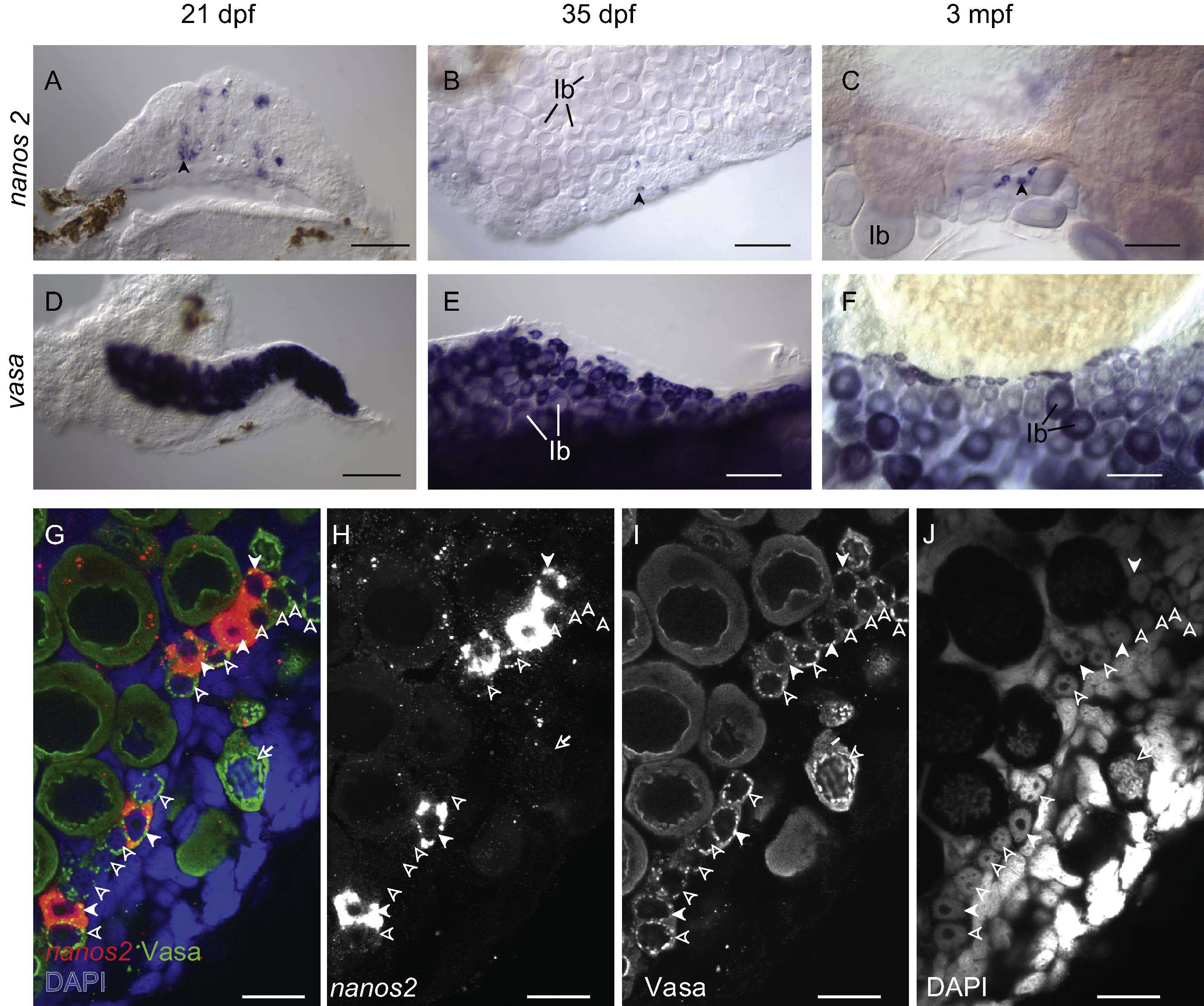
Fig. 3 nanos2 is expressed in a small subset of pre-meiotic oogonia. (A–C) RNA in situ hybridization shows that nanos2 (blue staining in A) is expressed in a subset of <20 μm germ cells, in comparison to vasa (blue staining in D) that is expressed in all germ cells. While nanos2-expressing cells are initially randomly distributed at 21 dpf (A), they become restricted to the lateral edges of the ovary by about 35 dpf (B). In adult ovaries, nanos2 continues to be expressed in a subset of <20 μm germ cells (C) as compared to vasa, which is expressed in all germ cells (F). Arrows in A–C identify single nanos2-expressing cells. (G–J) Fluorescent in situ hybridization for nanos2 mRNA and immunohistochemistry for Vasa protein (G) confirms that all nanos2-expressing cells (red in G; white in H) are germ cells, as they also express Vasa (green in G; white in I). Nuclear morphology, as revealed by DAPI staining (blue in G; white in J) indicates that nanos2 is expressed in a subset of pre-meiotic oogonia, which have characteristic uncondensed chromatin and prominent nucleoi. In G–J, nanos2-expressing cells, nanos2-negative oogonia, and meiotic oocytes, are indicated by filled arrowheads, empty arrowheads, and arrows, respectively. All images are from whole mount preparations. Stage Ib oocyte (Ib); Scale bars: 100 μm (A–F), 20 μm (G–J).
Reprinted from Developmental Biology, 374(2), Beer, R.L., and Draper, B.W., nanos3 maintains germline stem cells and expression of the conserved germline stem cell gene nanos2 in the zebrafish ovary, 308-318, Copyright (2013) with permission from Elsevier. Full text @ Dev. Biol.