
|
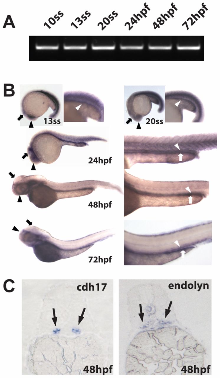
Fig. 3 Endolyn is expressed in the zebrafish kidney, brain and digestive system. (A) RT-PCR analysis of RNA extracted from embryos at the indicated stages was performed using specific primers against endolyn. All lanes show a band of the expected size (~600 bp). (B) In situ hybridization for endolyn in embryos was performed at 13ss and 20ss and at 24, 48, 72 hpf. Kidney (white arrowhead), brain (black arrow) and eye (black arrowhead) staining are evident by the 13ss stage and staining of the digestive system (white arrow) appears by 24 hpf. (C) Cross sections through the proximal tubule of zebrafish embryos at 48 hpf (5µm) were stained by in situ hybridization using probes to cadherin 17 (cdh17), a pronephric marker, or endolyn. Endolyn staining coincided with that of cadherin 17 in the expected region, confirming endolyn localization in the pronephric kidney.