
|
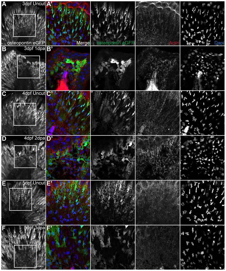
Fig. 7 The shape modification of mesenchymal cells lasts throughout and is specific of regeneration.
A,C,E Representative immunostaining with anti-GFP antibody in uncut transgenic osteopontin:eGFP larvae of 3 dpf, 4 dpf and 5 dpf respectively. B,D,F Representative immunostaining with anti-GFP antibody in amputated transgenic osteopontin:eGFP larvae of 3 dpf 1 dpa, 4 dpf 2 dpa and 5 dpf 3 dpa respectively. A–F Representative z-stack projections of the osteopontin:eGFP labeling. A′–F′ Representative single frames of the corresponding zoomed area represented by a square in A–F. Merged and single color images of osteopontin:eGFP labeling the mesenchymal cells (anti-GFP, green), actin (phalloidin, red) and nuclei (DAPI, blue), respectively. 5 Larvae per condition. Scale bar corresponds to 50 μm in all images.