
|
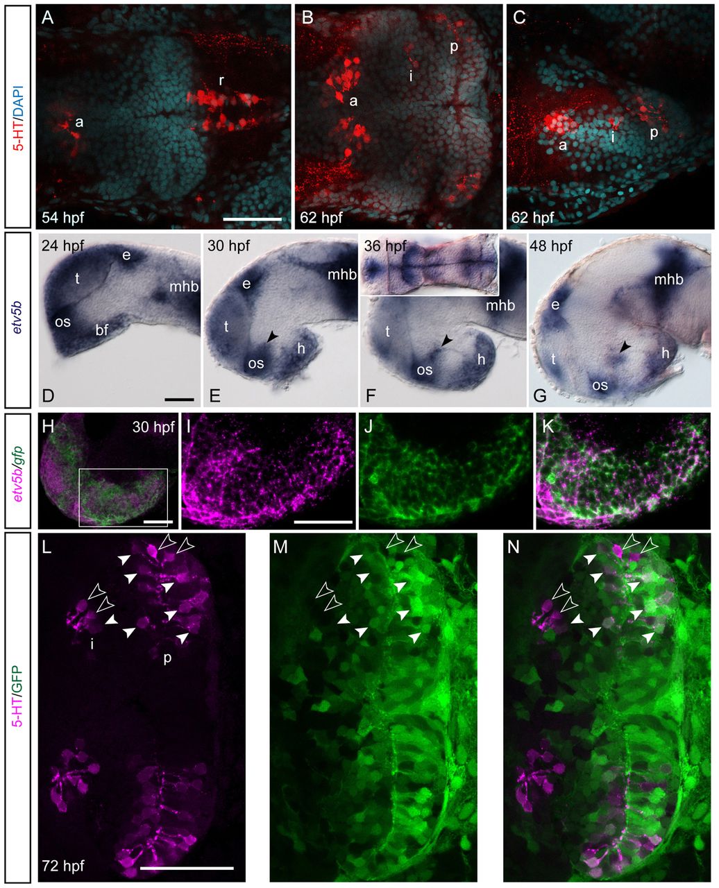
Fig. 1 5-HT precursors of the embryonic hypothalamus express etv5b. (A-C) Confocal maximum intensity projections of brains from zebrafish embryos processed for 5-HT immunohistochemistry and counterstained with DAPI (confocal optical section). Anterior (a.) and intermediate/posterior (i./p.) 5-HT clusters in the basal forebrain and the raphe population (r.) are indicated. (A,B) Ventral views; (C) lateral view, anterior left. (D-G) Expression of etv5b during embryonic development. etv5b transcripts were detectable in the basal forebrain (bf), at later stages including the hypothalamus (h) and a restricted region of the posterior tuberculum/hypothalamus (arrowheads). Further, transcripts were present in the pineal/epithalamus (e), telencephalon (t), optic stalk (os) and at the midbrain-hindbrain boundary (mhb). Lateral views, anterior left. Inset in F, ventral view. (H) Confocal maximum intensity projection of a Tg(ermp:gv)×Tg(uas:gfp) 30-hpf embryo processed for double gfp/etv5b ISH. Lateral view, anterior left. (I-K) High magnifications of boxed area in H; confocal optical sections. (L-N) Confocal optical section of brain from a Tg(ermp:gv)×Tg(uas:gfp) larvae revealing co-expression of GFP and 5-HT in neurons of the i./p. clusters. Ventral view, anterior left. White arrowheads indicate double-positive cells; open arrowheads indicate cells that are 5-HT-positive only. Scale bars: 50 μm.