
|
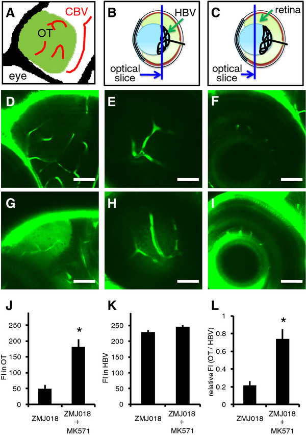
Fig. 4 Effect of MK571 on the permeability of the BBB and BRB to ZMJ018. Zebrafish larvae (albino line) at 7-8 dpf were immersed in egg water containing 1 μM of ZMJ018 with and without 30 μM of MK571 for 4 h. A-C: Schematic diagram showing the regions observed using a CLSM. D-I: In vivo fluorescence imaging of the OT (D and G), HBV (E and H) and multiple layers of the retina (F and I) in zebrafish stained with ZMJ018 only (D-F) or ZMJ018 in the presence of MK571 (G-I). The OT and multiple layers of the retina were clearly visible in zebrafish stained with ZMJ018 in the presence of MK571. J-L: Quantitative analysis of the FI in the OT (J) and HBV (K), and the ratio of the FI (L). Both the FI in the OT and the relative FI (OT/HBV) were significantly higher in zebrafish stained with ZMJ018 in the presence of MK571 (n = 4, *P < 0.05). Scale bar: 50 μm. OT, optic tectum; CBV, cerebral blood vessel; HBV, hyaloid blood vessel; FI, fluorescence intensity.