
Image
|
Figure Caption
Fig. S2
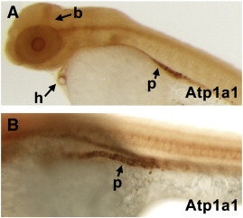
Localization of Atp1a1 protein. (A) Lateral view of Atp1a1 protein in 2 days post fertilization embryo. Atp1a1 protein is detected in the brain (b), heart (h), and pronephros (p). (B) A magnified image of the pronephros expression (p) detected by the anti-Atp1a1 antibody.
Figure Data
Acknowledgments
This image is the copyrighted work of the attributed author or publisher, and
ZFIN has permission only to display this image to its users.
Additional permissions should be obtained from the applicable author or publisher of the image.
Reprinted from Developmental Biology, 362(2), Langenbacher, A.D., Huang, J., Chen, Y., and Chen, J.N., Sodium pump activity in the yolk syncytial layer regulates zebrafish heart tube morphogenesis, 263-270, Copyright (2012) with permission from Elsevier. Full text @ Dev. Biol.