
|
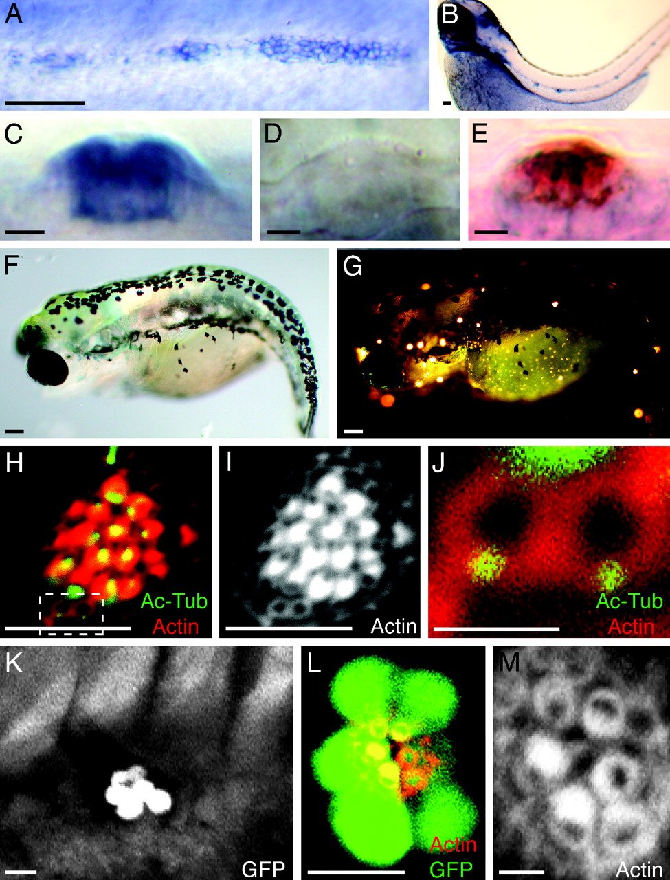
Fig. 3
Random orientation of hair bundles in neuromasts of trilobite mutant zebrafish. (A) In situ hybridization shows that vangl2 is expressed by the migrating primordium of the posterior lateral line in a 2-day-old wild-type embryo. (B) Expression is apparent in mature neuromasts at 3 days of age. (C) In a lateral view of a mature neuromast, the labeling indicates that the vangl2 transcript is expressed strongly by most neuromast cells. (D) A trilobitevu7 deletion mutant lacks vangl2 labeling. (E) A two-color in situ hybridization for vangl2 (blue) and the hair-cell marker parvalbumin 3a (red) indicates that vangl2 is expressed by both hair cells and supporting cells. (F) A brightfield micrograph displays the shortened, curled body of a trilobitem209 mutant larva at 5 days of age. (G) Fluorescence microscopy after exposure to 4-Di-2-Asp reveals that the trilobite larva possesses functional neuromasts along both the anterior and the posterior branches of its lateral-line system. (H–J) A neuromast of a 5-day-old trilobitem209 larva labeled for acetylated α-tubulin (green) and actin (red) shows that hair bundles assume random orientations. Two young hair cells occur at the lower edge of the neuromast (dashed box). (I) Staining of actin alone accentuates the defect in planar cell polarity. (J) A high-magnification view of the same neuromast reveals the mislocalization of basal bodies in two, probably sibling hair cells that have not yet formed mature hair bundles. (K) A low-magnification view of the trunk of an ET4;trilobite larva demonstrates that hair-cell pairs are aligned along a single axis in a neuromast. (L) A high-magnification view of a regenerating neuromast in an ET4;trilobite animal expressing GFP (green) and stained for actin (red) shows the alignment of hair cells along the neuromast′s line of symmetry. (M) In a higher-magnification view of this neuromast, actin staining demonstrates that, although the hair bundles respect the line of symmetry, they are misoriented with respect to one another. (Scale bars: A, B, F, and G, 100 μm; C–E, H, I, K, and L, 10 μm; J and M, 2 μm.)