
|
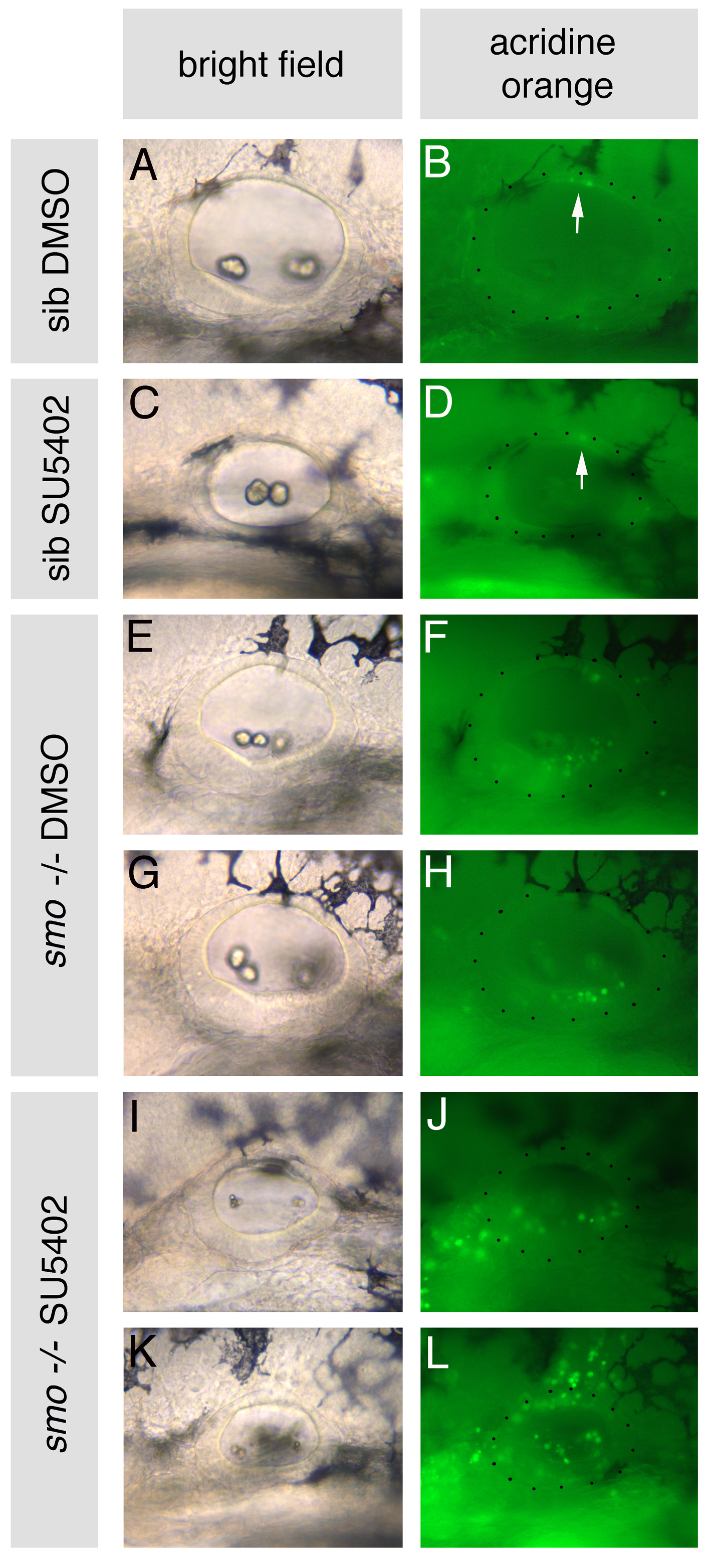
Fig. S6 Cell death is increased in otic vesicles of smo-/- embryos treated with SU5402. smo-/- and sibling (sib) embryos were treated with 10 μM SU5402 from 10 to 20S, stained with Acridine Orange for 1 hour between 38.5 and 39.5 hpf and imaged at 40 hpf. (A-D) Siblings treated with DMSO or SU5402 have negligible cell death in the otic vesicle; one or two dying cells are sometimes visible dorsally (arrow). (E-H) smo-/- embryos treated with DMSO display increased cell death in the otic vesicle ventrally, concentrated towards the posterior. (I-L) smo-/- embryos treated with SU5402 display increased cell death within the otic vesicle, at both the anterior and posterior, as well as around the otic vesicle. Lateral views; anterior to left, dorsal to top. Dots delineate the outline of the otic vesicle.