
|
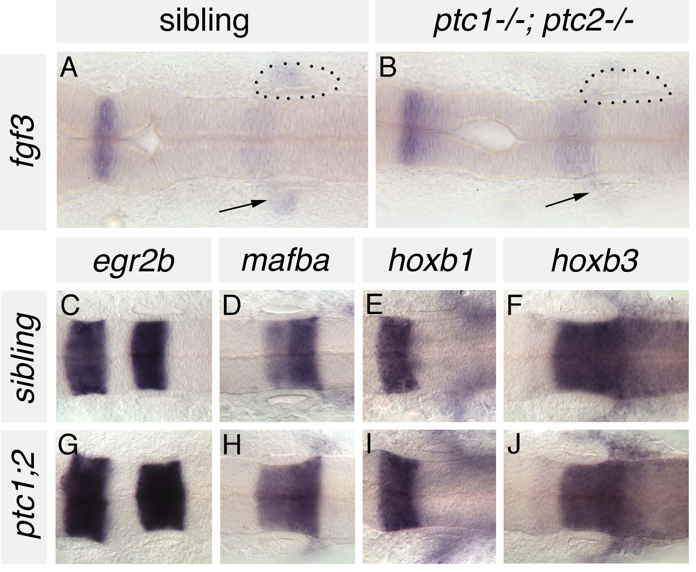
Fig. S5 Anteroposterior patterning is grossly normal in the hindbrain of ptc1-/-; ptc2-/- embryos. (A,B) In situ hybridisation to fgf3 in ptc1-/-; ptc2-/- embryos and wild-type siblings at 19S. Expression is reduced in the branchial arches (underneath the ear) of ptc1-/-; ptc2-/- embryos (arrows), but expressed at normal levels in rhombomere 4, adjacent to the anterior part of the developing inner ear. (C-J) In situ hybridisation to a panel of AP-restricted hindbrain markers in 22S ptc1-/-; ptc2-/- embryos and wild-type siblings. Expression of egr2b (krox20; rhombomeres 3 and 5), mafba (rhombomeres 5/6), hoxb1 (rhombomere 4) and hoxb3 (rhombomeres 5, 6 and more posteriorly) is grossly normal in the hindbrain of ptc1-/-;ptc2-/- embryos. Dorsal views; anterior to left. Dots delineate the otic vesicle in A and B.