
|
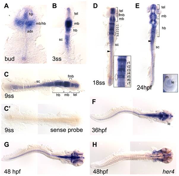
Fig. 2 her8a expression in the developing zebrafish embryos.
her8a expression is restricted in the developing nervous system during zebrafish embryogenesis analyzed by in situ hybridization. Stages of embryos shown in bottom left corner of each panel. Yolks were removed and embryos were flat-mounted, dorsal view except F and G lateral. her8a expression first appears in the developing brain at bud stage (A) and later becomes restricted to specific brain areas (B–E). The transcripts can be detected in the spinal cord from the 3-somite stage (3ss) (B) and retained until the latest stage analyzed (48 hpf) (C–G). Dashed circles in D mark the otic vesicles. Insert panel in D is an enlargement of the hindbrain, and in E is an enlargement of the eye. Arrowheads in D and E indicate her8a expressing cells at the midline. No signal was detected using sense riboprobe (C′). (H) her4 expression at 48 hpf . fmb, forebrain–midbrain boundary; hb, hindbrain; hp, hypothalamus; le, lens; mb, midbrain; r1-r7, rhombomere 1–7; rn, retinal neuroepithelial cells; sc, spinal cord; tel, telencephalon.