
|
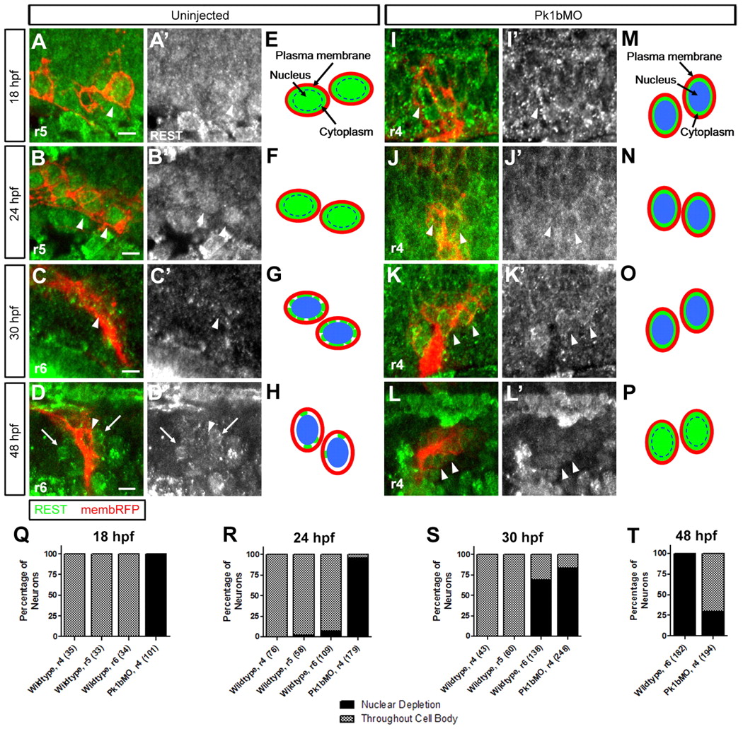
Fig. 6 REST is localized to the nuclei of migrating FBMNs. (A-D′,I-L′) Single-slice dorsal views of FBMNs (red) in Tg(zCREST1:membRFP) zebrafish embryos immunostained for REST (green) at the indicated stages. (A-D) In WT neurons, REST localized throughout the cell body during migration (A,B). As neurons settle in r6, REST becomes depleted from the nuclei (C). By 48 hpf, REST protein is further reduced or absent in FBMN cell bodies (D). (I-L) In Pk1b morphants, REST is depleted from the nuclei of FBMNs and expression is maintained at later embryonic stages. Arrowheads highlight individual FBMNs and arrows indicate non-FBMNs. A′-D′ and I′-L′ show REST immunostaining alone Scale bars: 10 μm. (E-H,M-P) Schematics illustrating REST localization (green) in FBMNs (red); nuclei are indicated (blue). See also Fig. S6 in the supplementary material. (Q-T) Localization of REST was scored in WT and Pk1bMO FBMNs. Percentage of FBMNs with depleted nuclear REST or localization throughout the cell body is displayed. The number of neurons scored is indicated in parentheses.