
|
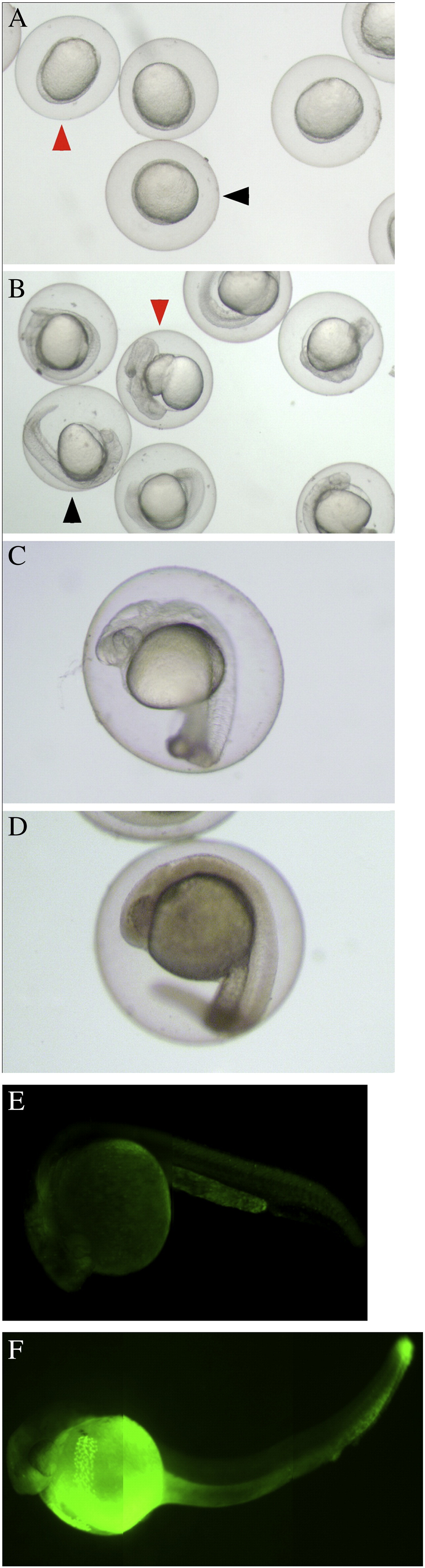
Fig. 4 Zebrafish embryos arrest development when exposed to 900 μM NaN3 in embryo medium. A) 6 hpf embryos were collected and exposed to 900 μM NaN3 in embryo medium for 24 h. B) Embryos were washed out of the NaN3 solution into embryo medium and left to resume development for an additional 24 h (Red arrowheads indicate typical abnormal embryos. Black arrowheads indicate normal embryos). C) An embryo at 24 hpf before exposure to 900 μM NaN3. D) An embryo after exposure to 900 μM NaN3 for 24 h. Note the brownish tinge and reduced transparency. However, such embryos are able to resume apparently normal development. E and F) Acridine orange staining to examine cell death. E) An embryo at 24 hpf that has developed under normoxia. F) An embryo that was allowed to develop to 24 hpf under normoxia before exposure to 900 μM NaN3 for 24 h. It was then washed three times for 5 min. in embryo medium before staining and observation.