
|
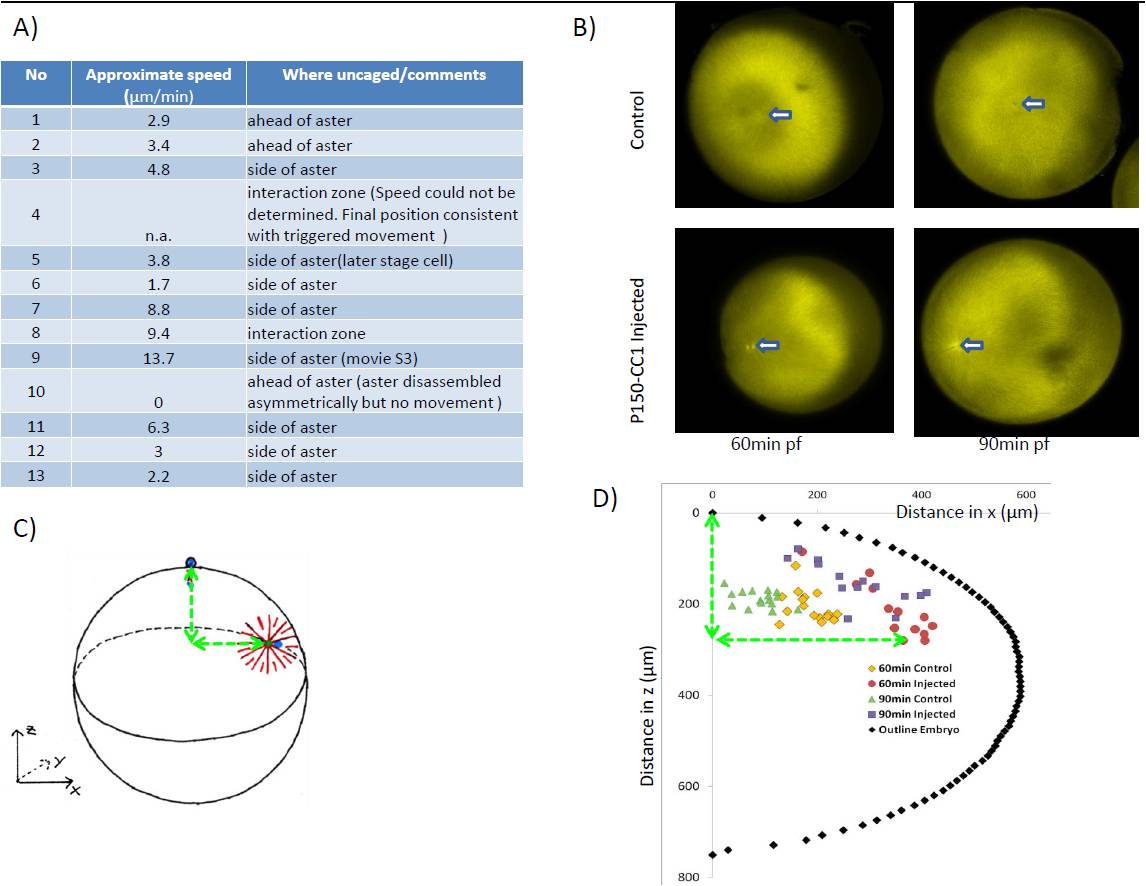
Fig. S3 Related to Figure 3. A) List of experiments in which microtubules were depolymerised locally in zebrafish embryos, together with the speed of aster movement. Speed was measured (approximately) by tracking the movement of fixed parts at the center of the aster (the centrosome or mid-body). Positive speeds indicate movement away from the irradiated zone. Movement towards the region of uncaging (which we did not observe) would have been reported as negative speed. In 12/13 experiments the aster moved away from the irradiated zone, in 1/13 it did not move and in no case was movement towards the zone observed. B-D) Sperm-aster in frog embryos depends on dynactin to move centrosome: Embryos were synchronously fertilized and fixed 60 and 90 min after fertilization. B) By staining against tubulin sperm aster formation and centrosome movement could be followed in p150-CC1 injected and control embryos. Arrow indicated position of centrosome. C) Because centrosome movement is three-dimensional, centrosome position was recorded relative to the animal pole at the top of the embryo. D) Centrosome position of injected and control embryos were plotted with circumference of one embryo. In control embryos centrosomes move from the periphery towards the cell’s center while p150-CC1 injected embryos centrosomes were located close to the cortex