
|
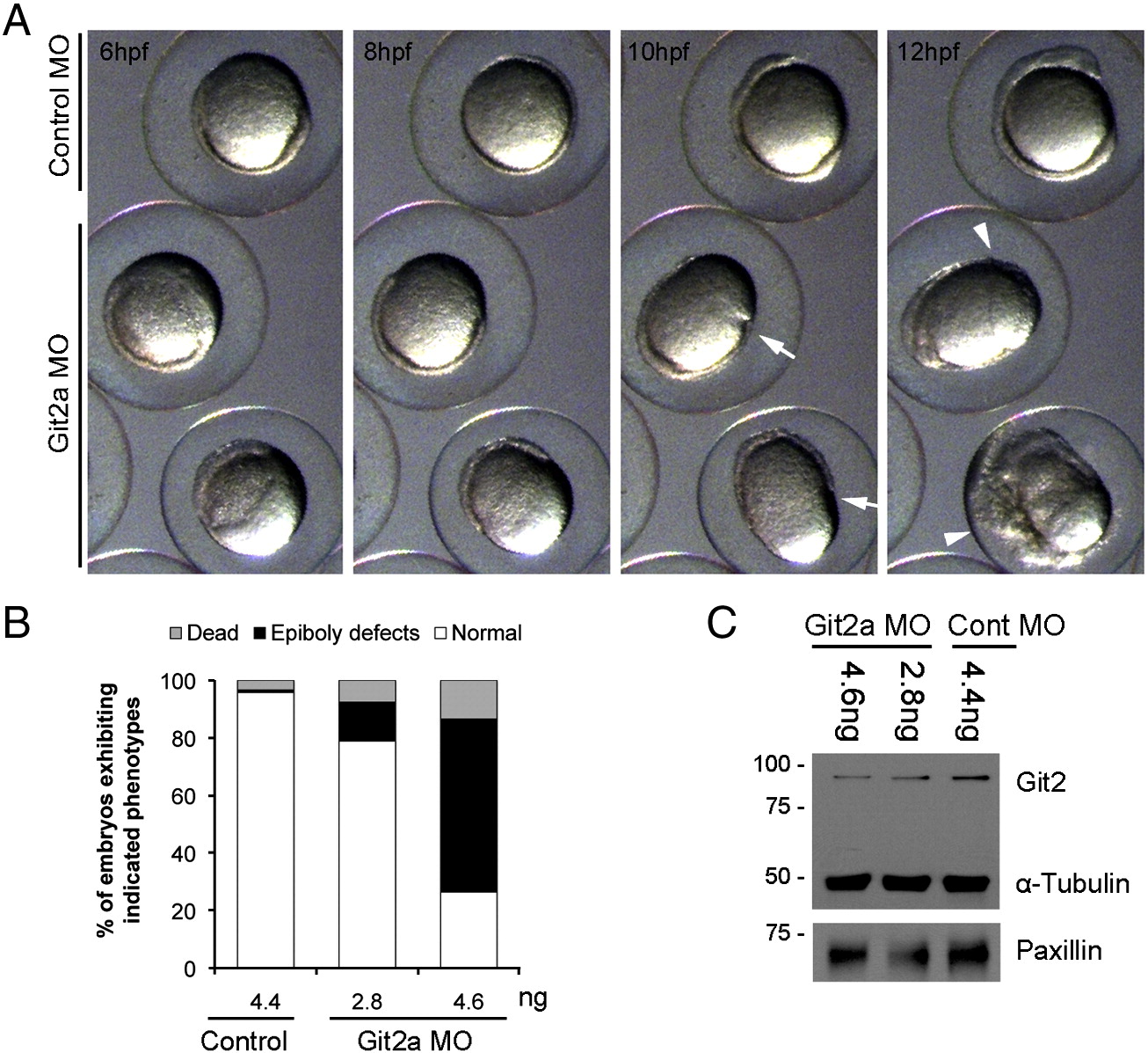
Fig. S3 Embryonic phenotypes of Git2a knockdown. (A) Montage of images from a representative time-lapse movie showing git2a morphant phenotypes at 6 hpf, 8 hpf, 10 hpf and 12 hpf. Constriction (white arrow) and cell movement delay or arrest (white arrowhead) was detected during epiboly in git2a morphants, which often resulted in embryonic lethality. (B) Quantification of epiboly defects in embryos examined at 9 hpf shows that the affect of git2a MO was dose-dependent. Data pooled from three independent experiments (n e 316 for each dose). (C) Western blotting of Git2 showing dose-dependent MO knockdown of Git2 protein.
Reprinted from Developmental Biology, 349(2), Yu, J.A., Foley, F.C., Amack, J.D., and Turner, C.E., The Cell Adhesion-associated Protein Git2 Regulates Morphogenetic Movements during Zebrafish Embryonic Development, 225-237, Copyright (2011) with permission from Elsevier. Full text @ Dev. Biol.