
|
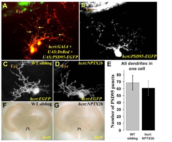
Fig. S6 Synapse distribution in HCRT dendrites is not affected by nptx2b overexpression. (A) The PSD95-EGFP postsynaptic fusion protein was visualized (yellow dots marked with arrows) on a background of cellular DsRed in dendrites of HCRT cells in 4dpf larvae. (B) To quantify synapse number in dendrites of single HCRT neurons, PSD95-EGFP was expressed transiently, and puncta were counted in the dendrites of 4dpf larvae. (E) At 4dpf, no significant differences were found in PSD95-EGFP puncta numbers between hcrt:NPTX2b and wild-type siblings. Statistical comparisons were performed using t-tests. Error bars indicate ± SEM. (C, D, F, G) Overexpression of nptx2b is not toxic to HCRT neurons, as visualization of EGFP indicates normal morphology in 4 dpf larvae (C, D) and adult hcrt:NPTX2b fish showed normal expression of hcrt mRNA (F, G).