
Image
|
Figure Caption
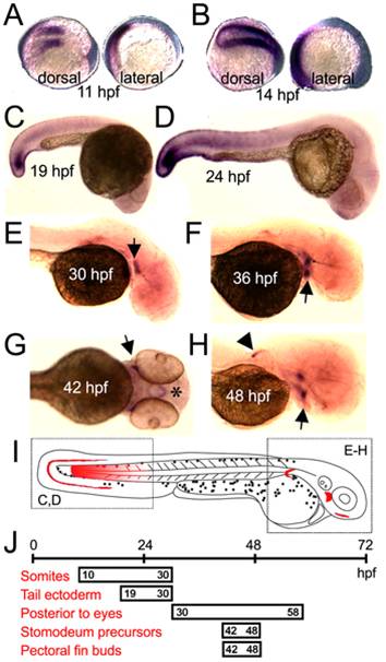
Fig. 5 Detailed analysis of CPA6 mRNA expression throughout zebrafish development.
In situ hybridization indicated CPA6 mRNA (purple) is found in newly formed somites (A–D), ectodermal cells of the tail (C,D), a tissue posterior to the eye (E–H, arrows), the stomodeum (G, asterisk), and the pectoral fins (H, arrowhead). CPA6 expression posterior to both left and right eyes can be seen in F and H. A summary of the spatial (I) and temporal (J) expression of CPA6 throughout zebrafish development is shown.
Figure Data
Acknowledgments
This image is the copyrighted work of the attributed author or publisher, and
ZFIN has permission only to display this image to its users.
Additional permissions should be obtained from the applicable author or publisher of the image.
Full text @ PLoS One