
|
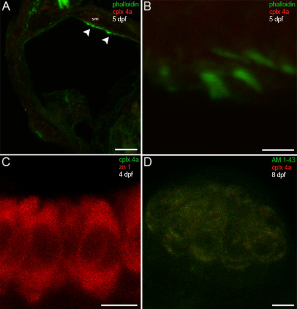
Fig. 10 Complexin 4a is not expressed in hair cells of the larval zebrafish acousticolateral system. (A) A low-magnification confocal micrograph of a transverse section through the otic vesicle of a 5-dpf zebrafish shows saccular macula (sm) inner ear hair cell stereocilia stained with phalloidin (green) that lack complexin 4a (red, arrowheads). (B) A high-magnification en face view of inner ear hair cells labeled with phalloidin (green) confirms the absence of complexin 4a (red) among their stereocilia. (C) Inner ear hair cells incubated with anti-zn 1 (red) lack complexin 4a (green). (D) A high-magnification confocal planar projection through a cranial neuromast labeled with AM1-43 (green) indicates that complexin 4a is also absent from these hair cells. Sections incubated with only secondary antibodies exhibit very low levels of diffuse immunofluorescence throughout the hair cells (data not shown). Scale bars: 25 μm (A); 5 μm (B-D).