
|
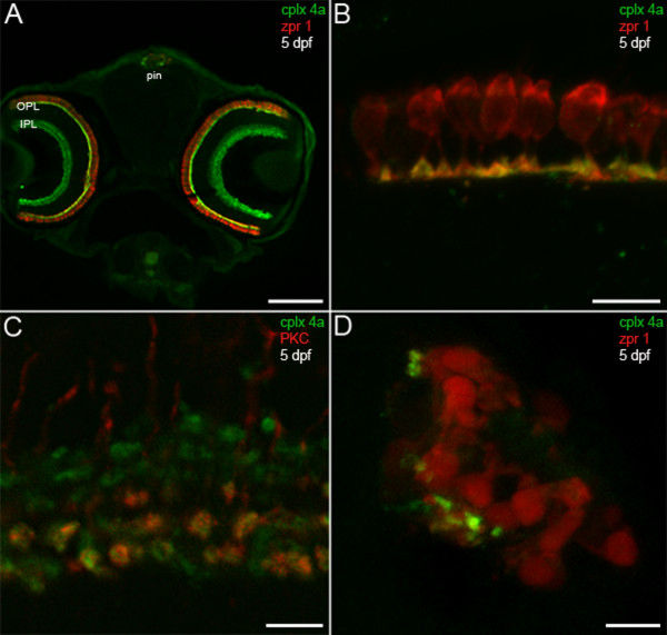
Fig. 9 Complexin 4a marks visual system ribbon presynaptic terminals. (A) A confocal micrograph of a 5-dpf zebrafish transverse section stained with the anti-complexin 4a antibody (green) and anti-zpr 1/FRet 43 (red). Note the similarity between the complexin 4a expression pattern and that of complexin 3/4 (Figure 3A) at low magnification in the retinal plexiform layers (OPL, IPL) and pineal organ (pin). (B) At high magnification, complexin 4a appears to be restricted primarily to the terminals of double cone photoreceptors in the retinal outer plexiform layer. (C) A confocal planar projection of the retinal inner plexiform layer shows overlap of complexin 4a (green) in protein kinase C (PKC)-positive (red) ON bipolar cell terminals in the IPL. (D) A confocal planar projection of a 5-dpf zebrafish sagittal section through the pineal organ is shown. Zpr 1/FRet 43-positive photoreceptors (red) are oriented such that their outer segments are medial and their short axons and presynaptic terminals are lateral. Complexin 4a (green) specifically localizes to putative terminals. Sections incubated with secondary antibodies alone exhibit background immunofluorescence in the retina and pineal (data not shown). Scale bars: 125 μm (A); 10 μm (B, D); 5 μm (C).