
|
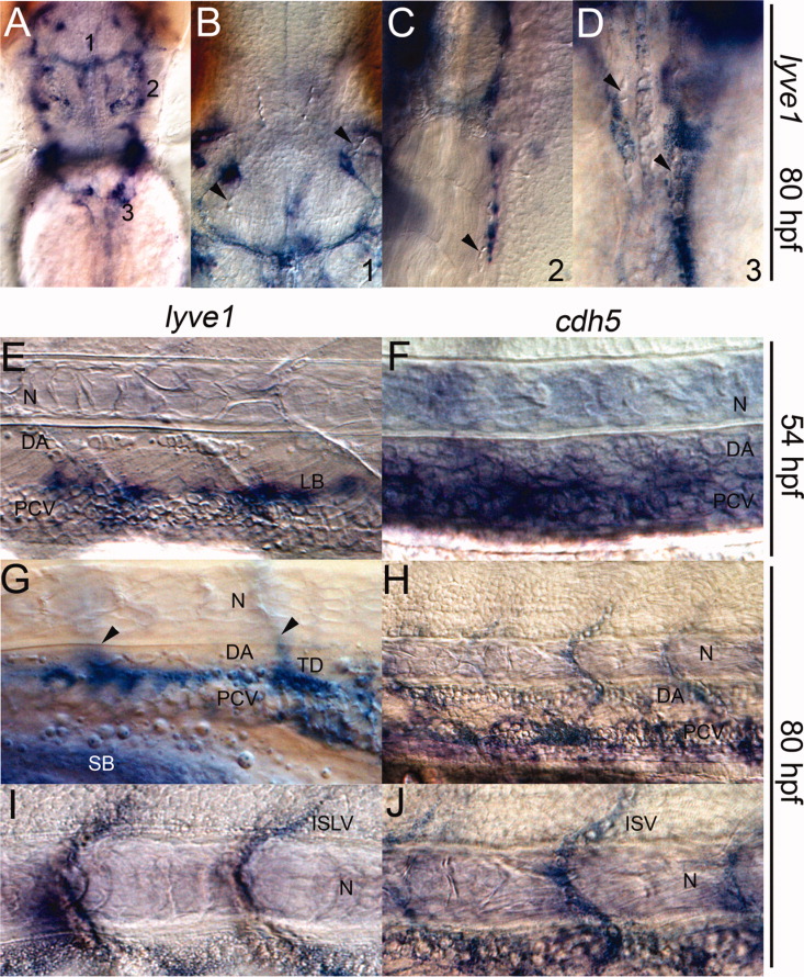
Fig. 3 Vessels expressing lyve1 do not support circulating blood. A-J: Nomarski image of the head; dorsal view (A-D) and trunk, lateral view (E-J). B,C,D: lyve1-expressing structures within the head do not contain erythrocytes (arrowheads). E,F: At 54 hours postfertilization (hpf): lymphangioblasts (LB) expressing lyve1 lay just above the blood-filled posterior cardinal vein (PCV) in E; cdh5 marked both the dorsal aorta (DA) and PCV in F. G-J: At 80 hpf: lyve1 expression within the developing thoracic duct (TD) and intersegmental lymphatic vessels (arrowheads, out of focus) in G; lyve1 marked intersegmental lymphatic vessels (ISLV) in I; cdh5 marked the blood-filled DA, PCV and intersegmental vessels (ISV) in H and J; N, notochord.