
|
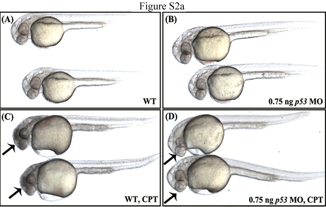
Fig. S2a The brain degeneration phenotype obtained for eif3ha morphants is likely not due to off-target effects mediated by up-regulation of p53. Panel a. Panels A and B represent wild-type uninjected embryos and embryos injected with 0.75 ng of p53-specific morpholino, respectively. Under these conditions, there is no apparent morphant phenotype. In panel C, wild-type 24 hours postfertilization (hpf) embryos were treated with 500 nM camptothecin (CPT) and cultured for 3-4 hr. This drug is known to induce p53-mediated apoptosis in zebrafish (Suppl. Ref: Langheinrich et al., 2002). The apoptosis induced by CPT is clearly visible in these embryos as dark tissue especially in the brain region (arrows). Panel D represents the p53-morphant embryos, which remained in CPT for 3-4 hr. In contrast to the wild-type embryos, CPT-mediated apoptosis was not observed (~95-100%, n > 30-40). This result shows that injection of 0.75 ng of this p53-specific morpholino is sufficient to rescue CPT-mediated apoptosis induced by up-regulation of p53.